

Roles of PPARγ/NF-κB signaling pathway in the pathogenesis of intrahepatic cholestasis of pregnancy
- PMID: 24489901
- PMCID: PMC3906154
- DOI: 10.1371/journal.pone.0087343
Roles of PPARγ/NF-κB signaling pathway in the pathogenesis of intrahepatic cholestasis of pregnancy
Abstract
Background: Intrahepatic cholestasis of pregnancy (ICP) is the most prevalent pregnancy specific liver disease. However, the pathogenesis and etiology of ICP is poorly understood.
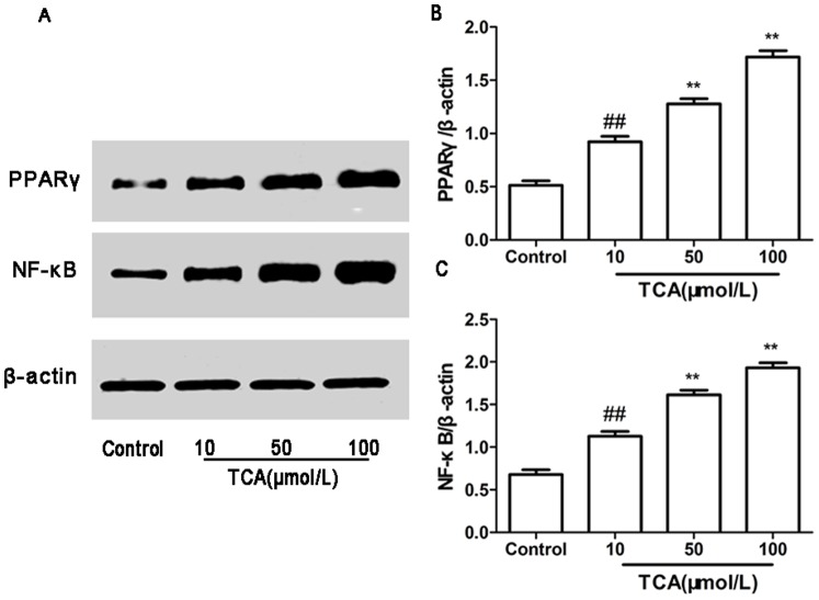
Aim: To assess the expression of peroxisome proliferator-activated receptorγ (PPARγ) and nuclear factor kappa B (NF-κB) in placenta and HTR-8/SVneo cell, and evaluate the serum levels of cytokines, bile acids, hepatic function and lipids in control and ICP patients and the fetal outcome, in order to explore the role of PPARγ/NF-κB signaling pathway in the possible mechanism of ICP.
Methods: Clinical data of the pregnant women were collected and serum levels of cytokines, bile acids, hepatic function and lipids were measured. Expressions of PPARγ and NF-κB in placenta and HTR-8/SVneo cell were determined. The new-born information was collected to demonstrate the relationship between PPARγ/NF-κB signaling pathway and ICP.
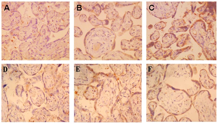
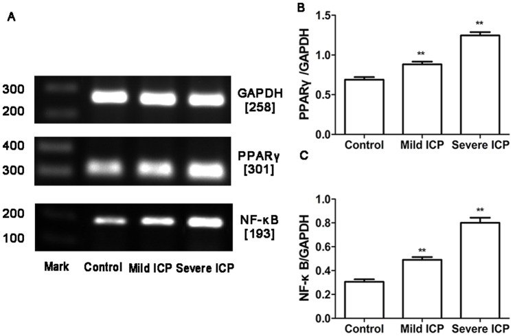
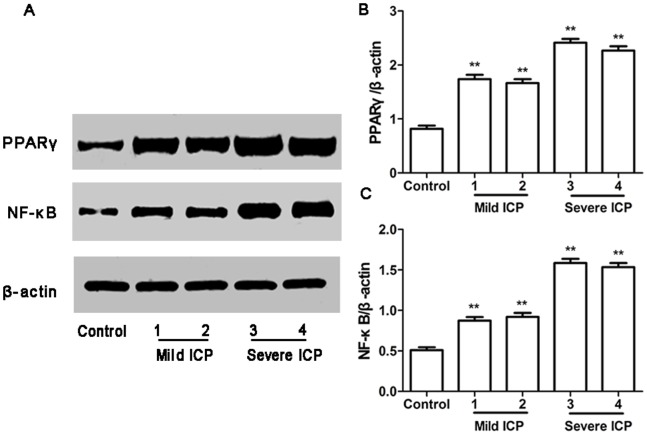
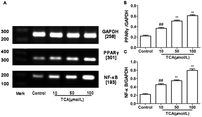
Results: The serum levels of bile acids, hepatic function, triglycerides (TG), total cholesterol (TC), IL-6, IL-12 and TNF-α in ICP group were significantly increased (P<0.01), and serum level of IL-4 was significantly decreased (P<0.01). PPARγ and NF-κB staining were found in the membrane and cytoplasm of placental trophoblast cell. The expression of PPARγ and NF-κB were significantly higher in ICP group and taurocholate acid (TCA) treated HTR-8/SVneo cell (P<0.01). The new-born information in severe ICP group were significantly different as compared to that in control group (P<0.05), and part of information in mild ICP group were also difference to that in control group (P<0.05).
Conclusions: The higher expressions of PPARγ and NF-κB in ICP placenta and TCA treated HTR-8/SVneo cell, together with the abnormal serum levels of cytokines, might induced by the imbalance of inflammatory and immune reaction, and then disturb placental bile acid and serum lipids transportation, finally result in fatal cholestasis which probably be one of the mechanism of ICP.
Conflict of interest statement
Figures
References
-
- Joshi D, James A, Quaglia A, Westbrook RH, Heneghan MA (2010) Liver disease in pregnancy. Lancet 375: 594–605. - PubMed
-
- Kenyon AP, Girling JC (2011) Obstetricscholestasis. RCOG green-top guidelines No 43. London: RCOG.
-
- Chen J, Deng W, Wang J, Shao Y, Ou M, et al. (2013) Primary bile acids as potential biomarkers for the clinical grading of intrahepatic cholestasis of pregnancy. Int J Gynaecol Obstet 122: 5–8. - PubMed
Publication types
MeSH terms
Substances
LinkOut - more resources
Full Text Sources
Other Literature Sources
Medical
Miscellaneous
