. 2014 Jan 28;9(1):e87385.
doi: 10.1371/journal.pone.0087385. eCollection 2014.

Rift Valley fever virus incorporates the 78 kDa glycoprotein into virions matured in mosquito C6/36 cells

Affiliations

Rift Valley fever virus incorporates the 78 kDa glycoprotein into virions matured in mosquito C6/36 cells

Hana M Weingartl et al. PLoS One. .

Abstract

Rift Valley fever virus (RVFV), genus Phlebovirus, family Bunyaviridae is a zoonotic arthropod-borne virus able to transition between distant host species, causing potentially severe disease in humans and ruminants. Viral proteins are encoded by three genomic segments, with the medium M segment coding for four proteins: nonstructural NSm protein, two glycoproteins Gn and Gc and large 78 kDa glycoprotein (LGp) of unknown function. Goat anti-RVFV polyclonal antibody and mouse monoclonal antibody, generated against a polypeptide unique to the LGp within the RVFV proteome, detected this protein in gradient purified RVFV ZH501 virions harvested from mosquito C6/36 cells but not in virions harvested from the mammalian Vero E6 cells. The incorporation of LGp into the mosquito cell line - matured virions was confirmed by immune-electron microscopy. The LGp was incorporated into the virions immediately during the first passage in C6/36 cells of Vero E6 derived virus. Our data indicate that LGp is a structural protein in C6/36 mosquito cell generated virions. The protein may aid the transmission from the mosquitoes to the ruminant host, with a possible role in replication of RVFV in the mosquito host. To our knowledge, this is a first report of different protein composition between virions formed in insect C6/36 versus mammalian Vero E6 cells.


Conflict of interest statement

Competing Interests: The authors have declared that no competing interests exist.

Figures

Figure 1
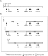
Figure 1. The M segment polyprotein.
Schematic summary of the viral proteins expressed from the M segment. Fig. 1.A. Translation from the M segment RNA can start at five start codons with methionine in amino acid (aa) positions 1, 39, 52, 131 and 136. All proteins are expressed using the same reading frame. The polyprotein has N-glycosylation sites at aa positions 88, 438, 794, 829, 1035 and 1077, and two cleavage sites between positions 153 and 154, and 690 and 691. The signal peptide (1–16 aa) is represented by a black thicker short line at the very N-terminus. Fig. 1.B. Different proteins are generated depending on the start codon used for the protein synthesis. Translation starting with methionine in position “1” yields LGp and Gc glycoproteins due to a cleavage at position 690/691. Both glycoproteins are fully glycosylated. Translation starting with methionine in aa position 39 yields three proteins: nonstructural NSm protein where the N-glycosylation site at aa position 88 may not be utilized, and two glycoproteins Gn and Gc due to cleavage in positions 153/154 and 690/691. No product has been identified for the putative starting methionine in position 52. Translation starting with methionine either at 131 or 136 position yields glycoproteins Gc and Gn, using the 690/691 cleavage site. Gn and Gc glycoproteins are considered to be fully glycosylated. Based on , , .
Figure 2
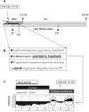
Figure 2. Selection of the peptide for antibody development.
Fig. 2.A. Schematic representation of the LGp/78 kDa glycoprotein (shaded bottom bar), Gn (gray top bar) and NSm (black striped bar) proteins. Gray full circles on stems represent the methionines in position 1 - start of the LGp/Gc polyprotein, and in position 39 - start of the NSm/Gn/Gc polyprotein. Forks indicate the two cleavage sites 153/154 and 690/691 in the M polyprotein. With translation starting at the methionine in position 39, cleavage at this sites leads to generation of the NSm, the Gn and the Gc proteins. With translation starting at the methionine in position 1, the cleavage occurs only at the 690/691 aa resulting in the LGp and Gc proteins. Clover leaves indicate the glycosylation sites (aa 88 and 438). Based on Gerrard and Nichol . Black solid bar represents the truncated recombinant recLGp (aa 1–121). Small thick fork indicates the peptide region unique to LGp against which the rabbit polyclonal (1109 and 1108) and the mouse monoclonal (SW9-22E) antibodies were raised. Fig. 2.B. Amino acid sequence of the recLGp including coding region of the expression vector at the N-terminus. Bold, capital M indicates starting methionine (V - in the expression plasmid, 1 - for LGp/Gc, 39 B for NSm/Gn/Gc);string of capital H stands for the His tag; underlined sequence from S to E in capital bold letters indicates sequence of the peptide used for antibody development. Italicized bold sequence dglnNit represents a potential prokaryotic N- glycosylation signal, and the eukaryotic N- glycosylation signal Nit (capital N in the 88 aa position). Fig. 2.C. Reprint of the EvoQuest predicted antigenicity of the SSTREETCTGDSTNPE peptide (the potential linear epitopes are encircled).
Figure 3
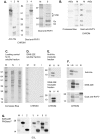
Figure 3. Characterization of antibodies.
Fig. 3.A. Left panel: Immunoblot of Sf9 cells lysates (soluble fraction) expressing baculovirus recombinant His-tagged Gn protein of RVFV (lanes 1 and 4) and bacterial cell lysate expressing the truncated recombinant LGp containing NSm sequence (lanes 2 and 3) detected with an anti-His antibody (lanes 1 and 2) or goat RVFV antiserum, (lanes 3 and 4). M – marker lane. CHROM  =  Chromogenic detection was used to detect antibody presence. Equal amount of protein (40 µg) was loaded per lane. Right panel, lane 5: Immunoblot of glycoproteins from purified RVFV produced in C6/36 cells using goat anti-RVFV serum. Fig. 3.B. Immunoblot of semipurified His tagged recombinant NSs protein of RVFV (400 ng of protein) detected with goat RVFV antiserum; M – marker lane. Left panel – Coomasie blue stained gel, right panel – immunoblot, chromogenic detection. Fig. 3.C. Confirmation of the specificity of the SW9-E22 antibody for the rLGp expressed in bacteria. Fig. 3.C is a loading control (Comassie Blue stained protein gel; PAGE was run using MES buffer) for the Fig. 3.D. M lane - protein marker, lane N - 40 µg of proteins from soluble fraction of the bacterial cell lysate, lane I – 40 µg of proteins from soluble fraction of the cell lysate from bacteria with IPTG induced protein synthesis. Fig. 3.D. Immunoblot of proteins in soluble fraction detected with SW9-E22 antibody against LGp or goat anti-mouse antibody conjugated with HRP using chromogenic visualization. Fig. 3.E. Immunoblot of the induced insoluble fraction from cell lysate of bacteria expressing the recLGp (lane I- I, 40 µg of protein) detected with anti-His antibody conjugated with HRP (top panel) or with SW9-22E antibody (bottom panel) using chromogenic detection. Lane N - transfected, uninduced E.coli B21 cell lysate (40 µg of protein). Lane M - protein size markers (SDS PAGE was run using MOPS buffer). Fig. 3.F. Immunoblot of the insoluble and soluble fractions from bacterial lysates expressing the truncated recombinant LGp (rLGp) with antibodies against the His tag detected with the mouse monoclonal antibody SW9-22E or with goat RVFV antiserum. M – marker lane, I-I induced insoluble fraction, I-S induced soluble fraction (20 µg of protein per lane), N - noninduced bacterial lysate (40 µg of protein). Fig. 3.G. Deglycosylation of the semi-purified recLGp detected with mouse SW9-E22 antibody and goat anti-mouse antibody conjugated with HRP uing chemiluminescent detection (ECL). Lane M - protein size markers (SDS PAGE was run using MES buffer); Lanes 1, 3 and 5 - untreated recLGp (400 ng); Lane 2, 4 and 6 - N-deglycosylated recLGp (400 ng), 24 hrs treatment with PNGase F.
Figure 4
Figure 4. Detection of LGp in cells infected with RVFV.
Fig. 4.A. Cell lysates of Vero E6 cells detected with the SW9-22E antibody using ECL detection. M- marker lane, C – uninfected cell control, 48 – cells infected with RVFV at 48 hpi. Protein loading 50 µg per lane. Fig.4 .B. Cell lysates of C6/36 cells detected with the SW9-22E antibody using ECL detection. M- marker lane, C – uninfected cell control, 96 – cells infected with RVFV at 96 hpi. Protein loading was 50 µg per lane. Black arrows indicate protein band expected to be the LGp; white arrow indicates a cellular protein upregulated during the RVFV infection. Fig. 4.C. Immunoblots of RVFV infected Vero E6 cells with goat RVFV antiserum using ECL detection. This membrane was stripped and re-probed with anti-actin antibody to confirm comparable protein loading in the individual lanes using the anti-actin antibody. M lane indicates the sizes of the protein markers. N lane - uninfected Vero E6 cell lysate negative controls. Lanes 24, 38 and 72 are RVFV infected Vero E6 cell lysates at 24, 48 and 72 hrs post infection (hpi). Protein loading was 50 µg per lane. Weaker detection of actin at 72 hpi is in agreement with expected block of cell protein synthesis during RVFV infection. Fig. 4.D. C6/36 cell control, mock infected at 96 h. Fig.4 .E. C6/36 cells infected with RVFV, 96 hpi. Magnification of 40× was used for both figures.
Figure 5
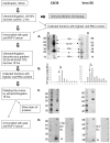
Figure 5. Summary of the virion purification process and an example of virion purification.
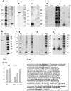
Purification flow of RVFV virions matured in C6/36 cells (second passage in C6/36 cells) is on the left panels of the figure; purification flow of the Vero E6 matured virions (first passage of C6/36 virus in Vero cells) is on the right panels of the figure. Fig. 5.A. Immunoblot using goat antiserum against RVFV of the fractions collected after concentration/semipurification through 20% sucrose onto 70% sucrose cushion. Beside structural proteins Gn/Gc and N, the LGp, as well as nonstructural proteins NSs and NSm were detected in the semipurified virion preparation. The assignment of RVFV proteins to protein bands reacting with the goat RVFV-antiserum was based on expected protein sizes, and known reactivity of the antiserum with respective recombinant proteins. Fig. 5.B. RVFV RNA profiles of fractions collected from the discontinuous 20 to 70% gradient (collected from the bottom). Fractions 10 and 11 were then pelleted down and lysed in the loading buffer for the SDS-PAGE. Fig. 5.C. Silver stain of the proteins from the gradient purified virion fractions separated by SDS-PAGE. Fig. 5.D. Aliquots of the samples from Fig.5.C. analyzed by immunoblotting using goat anti-RVFV serum (left panels designated L) or the SW9-22E antibody (right panels designated R). LGp was detected only in the C6/36 RVFV virions, both virion preparations had detectable levels of structural N and Gn/Gc proteins only.
Figure 6
Figure 6. Protein analysis of the gradient purified virion preparations.
Fig. 6.A. Preparation of RVFV virions grown in C6/36 cells (first passage of Vero E6 virus). 6.A.a Silver stained denaturing protein separation electrophoresis gel. 6.A.b Immunoblot of the same sample aliquot as in 6.A.a probed with the anti-LGp antibody SW9-22E. 6.A.c Immunoblot of the same membrane as in 6.A.b stripped, and re-probed with goat anti-RVFV serum. Fig.6 .A.d Immunoblot of purified RVFV virions matured in C6/36 cells probed with the antibody SW9-22E. Fig.6 .A.e Immunoblot of the same membrane as in 6.A.d stripped, and re-probed with goat anti-RVFV serum to illustrate that in independent samples, the monoclonal antibody SW9-E22 recognized only the smaller form of the LGp while the goat RVFV anti-serum detected only the larger form when used for re-probing the membranes. Fig. 6.B. Comparison of the C636 produced virions and the Vero E6 produced RVFV virions. Samples analyzed in Figure 6.A.a and in Figure 6.C.a (lane 1) were analyzed again on the same gel, and probed with anti-LGp antibody SW9-E22. C6 - RVFV virion preparation in C6/36 cells; V6 - RVFV virion preparation in Vero E6 cells. Fig. 6.C. Preparation of RVFV virions grown in Vero E6 cells (fourth passage). This preparation yielded double peak on the sucrose gradient, and the peaks were analyzed separately. Sample 1 are fractions collected at around 58% of sucrose and sample 2 at about 53% of sucrose. 6.C.a Silver stained denaturing protein separation electrophoresis gel. Lane designated as 1 is sample 1, lane 2 is sample 2. 6.C.b immunoblot of the same sample aliquots as in 6.C.a probed with anti-LGp antibody SW9-22E. 6.C.c immunoblot of the same membrane as in 6.C.b stripped, and re-probed with goat anti-RVFV serum. Protein separation gels for immunoblotting had both molecular size markers - colorimetric (Mc) and biotin-labeled (Me), and only the ladder closest to samples of interest is presented. Fig.6 .D.a Semi-quantification of the gel loading by measuring intensity of the band for the 28 kDa marker, and the band for the N protein on silver stained gels: A.a. protein densities for Fig.6.A.a - virions produced in C6/36 cells in. C.a protein densities for Fig.6.C.a – virions produced in Vero E6 cells. The intensity comparison indicates that the gels with Vero E6 produced virions were overloaded compare to the gels with C6/36 produced virions, considering that equal amounts of the marker proteins were used in all SDS-PAGE. Fig.6 .D.b Amino acid coverage of the LGp in bold letters as detected by mass spectrophotometry.
Figure 7
Figure 7. Visualization of the virions by electron miscroscopy.
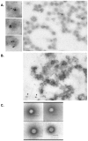
Fig. 7.A. Immune electron microscopy of virions produced in C6/36 cells labeled with mouse SW9-22E antibody tagged with 6 nm gold particles with detailed view of selected virions in separate panels. Fig. 7.B. Immune electron microscopy of virions produced in Vero E6 cells labeled with mouse SW9-22E antibody. Fig. 7.C. Negative staining of virions harvested from Vero E6 cells (upper panels) and negative staining of virions harvested from C6/36 cells (bottom panels). Scale bar represents 100 nm.

Similar articles

Cited by

References

    1. Pepin M, Bouloy M, Bird BH, Kemp A, Paweska J (2010) Rift Valley fever virus (Bunyaviridae: Phlebovirus): an update on pathogenesis, molecular epidemiology, vectors, diagnostics and prevention. Vet Res 41: 61 10.1051/vetres/2010033 - DOI - PMC - PubMed
    1. Collett MS, Purchio AF, Keegan K, Frazier S, Hays W, et al. (1985) Complete nucleotide sequence of the M RNA segment of Rift Valley fever virus. Virology 144: 228–245. - PubMed
    1. Torborg Kakach L, Suzich JA, Collett MS (1989) Rift Valley fever virus M segment: Phlebovirus expression strategy and protein glycosylation. Virology 170: 505–510. - PubMed
    1. Gerrard SR, Nichol ST (2007) Synthesis, proteolytic processing and complex formation of N-terminally nested precursor proteins of the Rift Valley fever virus glycoproteins. Virology 357: 124–133. - PMC - PubMed
    1. Gerrard SR, Bird BH, Albariño CG, Nichol ST (2007) The NSm proteins of Rift Valley fever virus are dispensable for maturation, replication and infection. Virology 359: 459–65. - PMC - PubMed

Publication types

LinkOut - more resources