. 2014 Jan 15;6(2):119-28.
eCollection 2014.

Transplantation of Fas-deficient or wild-type neural stem/progenitor cells (NPCs) is equally efficient in treating experimental autoimmune encephalomyelitis (EAE)

Affiliations

Transplantation of Fas-deficient or wild-type neural stem/progenitor cells (NPCs) is equally efficient in treating experimental autoimmune encephalomyelitis (EAE)

Charles Hackett et al. Am J Transl Res. .

Abstract

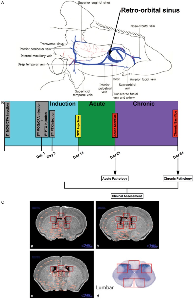
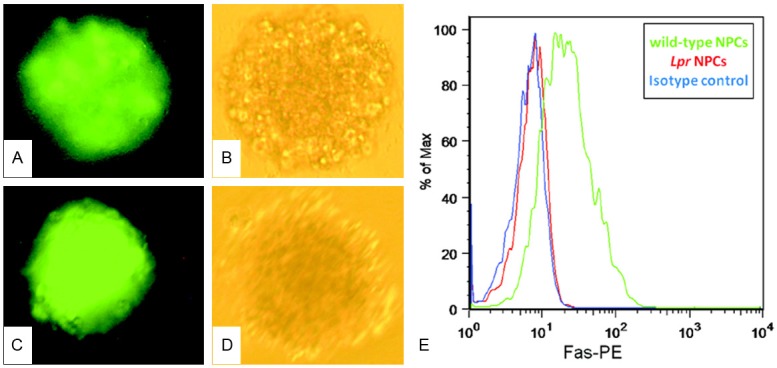
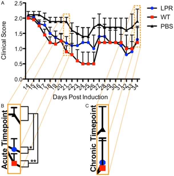
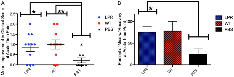
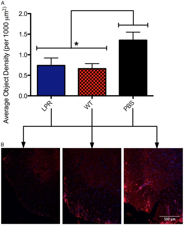
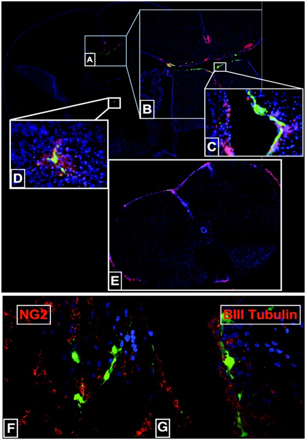
Studies have shown that neural stem/progenitor cell (NPC) transplantation is beneficial in experimental autoimmune encephalomyelitis (EAE), an established animal model of multiple sclerosis (MS). It is unclear whether NPCs have the ability to integrate into the host CNS to replace lost cells or if their main mechanism of action is via bystander immunomodulation. Understanding the mechanisms by which NPCs exert their beneficial effects as well as exploring methods to increase post-transplantation survival and differentiation is critical to advancing this treatment strategy. Using the EAE model and Fas-deficient (lpr) NPCs, we investigated the effects of altering the Fas system in NPC transplantation therapy. We show that transplantation of NPCs into EAE mice ameliorates clinical symptoms with greater efficacy than sham treatments regardless of cell type (wt or lpr). NPC transplantation via retro-orbital injections significantly decreased inflammatory infiltrates at the acute time point, with a similar trend at the chronic time point. Both wt and lpr NPCs injected into mice with EAE were able to home to sites of CNS inflammation in the periventricular brain and lumbar spinal cord. Both wt and lpr NPCs have the same capacity for inducing apoptosis of Th1 and Th17 cells, and minimal numbers of NPCs entered the CNS. These cells did not express terminal differentiation markers, suggesting that NPCs exert their effects mainly via bystander peripheral immunomodulation.

Keywords: Experimental autoimmune encephalomyelitis (EAE); Fas-deficient (lpr) NPC; neural progenitor cells (NPC); neuroprotection.


Figures

Figure 1
Figure 1
EAE induction protocol and experimental design. A: Map of blood flow in the mouse head along with injection site of the retro-orbital sinus of the adult mouse [19]. B: Timeline of experimental protocol illustrating induction procedures, treatment time-point, and clinico-pathological chronology. C: Templates used for image capture and quantification. a-c: represent the three periventricular brain depths (anterior to posterior) used, while d represents a lumbar spinal section. Red squares on the sections denote image locations.
Figure 2
Figure 2
Characterization of NPCs in vitro. (A) Lpr GFP+ NPCs form neurosphere and auto-fluoresce green under fluorescence. Images (20x) of 10-day-old GFP+ neurospheres grown in culture. GFP+ NPCs fluorescing green, with (B) a phase contrast image of the same sphere. (C) Wt GFP+ NPCs fluorescing green with (D) a phase contrast image of the same sphere. (E) Confirmation that Lpr NPCs lack FasR. Lpr mice have a large insertion in the FasR gene that results in improper splicing and absence of the protein. We confirmed absence of FasR in lpr NPC using a phycoerythrin (PE)-conjugated anti-FasR antibody. Background staining was assessed using an isotype control IgG antibody (blue line). Unlike wild-type NPCs (green line), lpr NPCs (red line) do not express FasR (+ peak completely overlaps with isotype control). Y-axis represents the percent of maximum staining intensity (arbitrary units), while the X-axis represents the voltage of positive PE fluorophore staining.
Figure 3
Figure 3
Wt and lpr NPC transplantation decreases EAE clinical scores. A: Mean clinical scores from each day after injection (error bars represent SEM). B: Mean clinical scores at the day of harvesting at the acute time point day 21 for lpr (n=11, p≤0.05), wt (n=10, p≤0.01) and PBS (n=9) treated mice. C: Mean clinical scores at the day of harvesting at the chronic time point day 34 for lpr (n=5), wt (n=5), and PBS (n=5) treated mice.
Figure 4
Figure 4
Wt and lpr NPCs have the same efficacy in EAE. A: Mean improvement in clinical score for lpr (n=11, p≤0.05) wt (n=10, p≤0.01) and PBS treated mice at the acute time point. Improvement in clinical score was determined by subtracting the clinical score at sacrifice from the clinical score at NPC injection (error bars represent SEM). B: Percent of mice showing recovery at acute time point for lpr (n=11, p≤0.05), wt (n=10) and PBS (n=9). A mouse was considered to be showing recovery if the clinical score at the time of sacrifice was less than the clinical score at the time of NPC injection (error bars represent SEM).
Figure 5
Figure 5
Wt and lpr NPCs decrease inflammation in EAE. A: Mean average object density of CD-45 positive cells in lpr (n=6, p≤0.05), wt (n=4, p≤0.05), and PBS determined using Neurolucida image processing software (error bars represent SEM). B: Representative 20x images of lumbar spinal sections of lpr (left), wt (middle), and PBS (right) stained for CD-45 positive cells (red cells).
Figure 6
Figure 6
NPCs home to the CNS and reside there at the acute and chronic time points. A: 5x image of transplanted NPCs (green cells) and CD-45 positive cells (red cells) in the periventricular region of the brain. B-D: Close-up images of the periventricular brain at 5x, 40x, and 40x respectively. E: 5x compilation image of the lumbar spine of a mouse transplanted with NPCs. F: Transplanted NPCs express a negligible amount of oligoprogenitor and neuroprogenitor cell lineage markers at the acute and chronic time points. (left) Image of a periventricular brain section stained for NG2 (an oligoprogenitor cell marker) in red and transplanted NPCs in green. (right) 60x image of a periventricular brain section stained for BIII Tubulin (a neuron specific cell marker) in red and transplanted NPCs in green.

Similar articles

Cited by

References

    1. Martino G, Pluchino S. The therapeutic potential of neural stem cells. Nat Rev Neurosci. 2006;7:395–406. - PubMed
    1. Galli R, Gritti A, Bonfanti L, Vescovi AL. Neural stem cells: an overview. Circ Res. 2003;92:598–608. - PubMed
    1. Einstein O, Karussis D, Grigoriadis N, Mizrachi-Kol R, Reinhartz E, Abramsky O, Ben-Hur T. Intraventricular transplantation of neural precursor cell spheres attenuates acute experimental allergic encephalomyelitis. Mol Cell Neurosci. 2003;24:1074–1082. - PubMed
    1. Pluchino S, Quattrini A, Brambilla E, Gritti A, Salani G, Dina G, Galli R, Del Carro U, Amadio S, Bergami A, Furlan R, Comi G, Vescovi AL, Martino G. Injection of adult neurospheres induces recovery in a chronic model of multiple sclerosis. Nature. 2003;422:688–694. - PubMed
    1. Politi LS, Bacigaluppi M, Brambilla E, Cadioli M, Falini A, Comi G, Scotti G, Martino G, Pluchino S. Magnetic-resonance-based tracking and quantification of intravenously injected neural stem cell accumulation in the brains of mice with experimental multiple sclerosis. Stem Cells. 2007;25:2583–2592. - PubMed

LinkOut - more resources