

Development of a Comprehensive Osteochondral Allograft MRI Scoring System (OCAMRISS) with Histopathologic, Micro-Computed Tomography, and Biomechanical Validation
- PMID: 24489999
- PMCID: PMC3904392
- DOI: 10.1177/1947603513514436
Development of a Comprehensive Osteochondral Allograft MRI Scoring System (OCAMRISS) with Histopathologic, Micro-Computed Tomography, and Biomechanical Validation
Abstract
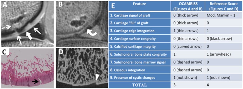
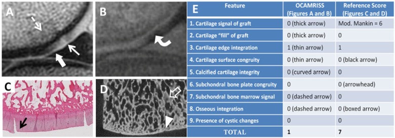
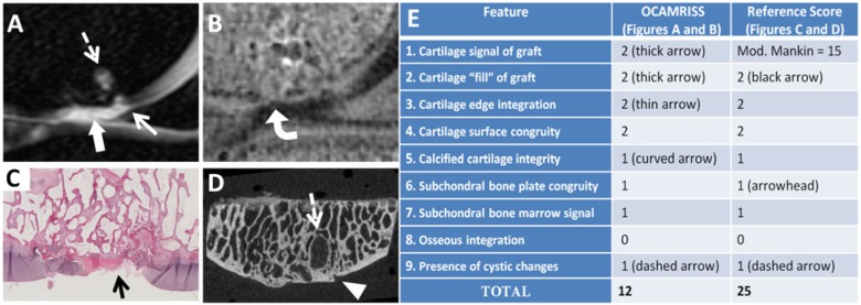
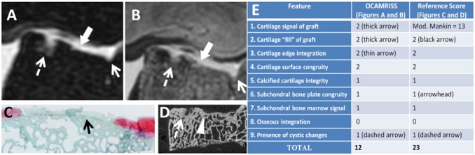
Objective: To describe and apply a semi-quantitative MRI scoring system for multi-feature analysis of cartilage defect repair in the knee by osteochondral allografts, and to correlate this scoring system with histopathologic, micro-computed tomography (μCT), and biomechanical reference standards using a goat repair model.
Design: Fourteen adult goats had two osteochondral allografts implanted into each knee: one in the medial femoral condyle (MFC) and one in the lateral trochlea (LT). At 12 months, goats were euthanized and MRI was performed. Two blinded radiologists independently rated nine primary features for each graft, including cartilage signal, fill, edge integration, surface congruity, calcified cartilage integrity, subchondral bone plate congruity, subchondral bone marrow signal, osseous integration, and presence of cystic changes. Four ancillary features of the joint were also evaluated, including opposing cartilage, meniscal tears, synovitis, and fat-pad scarring. Comparison was made with histological and μCT reference standards as well as biomechanical measures. Interobserver agreement and agreement with reference standards was assessed. Cohen's kappa, Spearman's correlation, and Kruskal-Wallis tests were used as appropriate.
Results: There was substantial agreement (κ>0.6, p<0.001) for each MRI feature and with comparison against reference standards, except for cartilage edge integration (κ=0.6). There was a strong positive correlation between MRI and reference standard scores (ρ=0.86, p<0.01). OCAMRISS was sensitive to differences in outcomes between the types of allografts.
Conclusions: We have described a comprehensive MRI scoring system for osteochondral allografts and have validated this scoring system with histopathologic and μCT reference standards as well as biomechanical indentation testing.
Keywords: MRI scoring system; cartilage repair; osteochondral allografts.
Conflict of interest statement
Figures


Similar articles
-
Osteochondral Allograft MRI Scoring System (OCAMRISS) in the Knee: Interobserver Agreement and Clinical Application.Cartilage. 2015 Jul;6(3):142-9. doi: 10.1177/1947603515573987. Cartilage. 2015. PMID: 26175859 Free PMC article.
-
Bone Marrow Aspirate Concentrate Does Not Improve Osseous Integration of Osteochondral Allografts for the Treatment of Chondral Defects in the Knee at 6 and 12 Months: A Comparative Magnetic Resonance Imaging Analysis.Am J Sports Med. 2019 Feb;47(2):339-346. doi: 10.1177/0363546518813915. Epub 2018 Dec 13. Am J Sports Med. 2019. PMID: 30543757
-
Decreased Graft Thickness Is Associated With Subchondral Cyst Formation After Osteochondral Allograft Transplantation in the Knee.Am J Sports Med. 2019 Jul;47(9):2123-2129. doi: 10.1177/0363546519851098. Epub 2019 Jun 6. Am J Sports Med. 2019. PMID: 31169995
-
Autologous tissue transplantations for osteochondral repair.Dan Med J. 2016 Apr;63(4):B5236. Dan Med J. 2016. PMID: 27034191 Review.
-
Role of Particulated Juvenile Cartilage Allograft Transplantation in Osteochondral Lesions of the Talus: A systematic review.Foot Ankle Surg. 2021 Jan;27(1):10-14. doi: 10.1016/j.fas.2020.02.011. Epub 2020 Feb 26. Foot Ankle Surg. 2021. PMID: 32169329
Cited by
-
Return to Play Among Elite Basketball Players After Osteochondral Allograft Transplantation of Full-Thickness Cartilage Lesions.Orthop J Sports Med. 2018 Jul 25;6(7):2325967118786941. doi: 10.1177/2325967118786941. eCollection 2018 Jul. Orthop J Sports Med. 2018. PMID: 30109237 Free PMC article.
-
A new computed tomography scoring system to assess osteochondral allograft transplantation for the knee: inter-observer and intra-observer agreement.Int Orthop. 2021 May;45(5):1191-1197. doi: 10.1007/s00264-020-04927-w. Epub 2021 Jan 8. Int Orthop. 2021. PMID: 33416905
-
Evaluation of Dexamethasone-Eluting Cell-Seeded Constructs in a Preclinical Canine Model of Cartilage Repair.Tissue Eng Part A. 2025 Feb;31(3-4):208-218. doi: 10.1089/ten.tea.2024.0244. Epub 2024 Nov 28. Tissue Eng Part A. 2025. PMID: 39607494
-
Osteochondral allograft transplantation in cartilage repair: Graft storage paradigm, translational models, and clinical applications.J Orthop Res. 2016 Jan;34(1):31-8. doi: 10.1002/jor.22998. Epub 2015 Sep 24. J Orthop Res. 2016. PMID: 26234194 Free PMC article. Review.
-
Sustained low-dose dexamethasone delivery via a PLGA microsphere-embedded agarose implant for enhanced osteochondral repair.Acta Biomater. 2020 Jan 15;102:326-340. doi: 10.1016/j.actbio.2019.11.052. Epub 2019 Dec 2. Acta Biomater. 2020. PMID: 31805408 Free PMC article.
References
-
- Curl WW, Krome J, Gordon ES, Rushing J, Smith BP, Poehling GG. Cartilage injuries: a review of 31,516 knee arthroscopies. Arthroscopy. 1997;13:456-60. - PubMed
-
- Robertsson O, Dunbar M, Pehrsson T, Knutson K, Lidgren L. Patient satisfaction after knee arthroplasty: a report on 27,372 knees operated on between 1981 and 1995 in Sweden. Acta Orthop Scand. 2000;71:262-7. - PubMed
-
- Hunziker EB. Articular cartilage repair: basic science and clinical progress. A review of the current status and prospects. Osteoarthritis Cartilage. 2002;10:432-63. - PubMed
-
- Delcogliano A, Caporaso A, Menghi A, Rinonapoli G, Chiossi S. Results of autologous osteochondral grafts in chondral lesions of the knee. Minerva Chir. 2002;57:273-81. - PubMed
-
- Jakob RP, Franz T, Gautier E, Mainil-Varlet P. Autologous osteochondral grafting in the knee: indication, results, and reflections. Clin Orthop Relat Res. 2002;(401):170-184. - PubMed
Grants and funding
LinkOut - more resources
Full Text Sources
Other Literature Sources
