. 2013:2013:637629.
doi: 10.1155/2013/637629. Epub 2013 Dec 30.

Genetics of proteasome diseases

Affiliations

Genetics of proteasome diseases

Aldrin V Gomes. Scientifica (Cairo). 2013.

Abstract

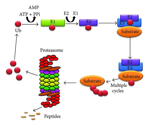
The proteasome is a large, multiple subunit complex that is capable of degrading most intracellular proteins. Polymorphisms in proteasome subunits are associated with cardiovascular diseases, diabetes, neurological diseases, and cancer. One polymorphism in the proteasome gene PSMA6 (-8C/G) is associated with three different diseases: type 2 diabetes, myocardial infarction, and coronary artery disease. One type of proteasome, the immunoproteasome, which contains inducible catalytic subunits, is adapted to generate peptides for antigen presentation. It has recently been shown that mutations and polymorphisms in the immunoproteasome catalytic subunit PSMB8 are associated with several inflammatory and autoinflammatory diseases including Nakajo-Nishimura syndrome, CANDLE syndrome, and intestinal M. tuberculosis infection. This comprehensive review describes the disease-related polymorphisms in proteasome genes associated with human diseases and the physiological modulation of proteasome function by these polymorphisms. Given the large number of subunits and the central importance of the proteasome in human physiology as well as the fast pace of detection of proteasome polymorphisms associated with human diseases, it is likely that other polymorphisms in proteasome genes associated with diseases will be detected in the near future. While disease-associated polymorphisms are now readily discovered, the challenge will be to use this genetic information for clinical benefit.


Figures

Figure 1
Figure 1
Schematic diagram of the ubiquitin-proteasome system. The UPS involves at least three enzymes (E1, E2, and E3) that catalyze the addition of ubiquitin to lysine residues on the substrate protein. Polyubiquitinated substrates are then recognized by the proteasome or proteasome associating protein, and the ubiquitin removed by deubiquitinases and the substrate unfolded and translocated in the 20S core for proteolysis.
Figure 2
Figure 2
Schematic diagram of the 26S proteasome. The 26S proteasome is composed of the core 20S proteasome and the 19S proteasome complex.
Figure 3
Figure 3
Schematic diagram of different forms of the proteasome. Intracellular proteasome exists in different forms. The 26S proteasome can exist with one or two 19S caps, immunoproteasomes containing one or two 11S caps, proteasomes containing the 20S proteasome with one or two PA200 caps (in the nucleus only), and hybrid proteasomes which contain different combinations of 20S and activators.
Figure 4
Figure 4
Alignment of human PSMA subunits 1–8. Protein sequences of the eight proteasome PSMA subunits were aligned using Clustal W. ⋆, identical residue in all seven subunits;  :, conserved amino acids with strongly similar properties; ., conservation between residues of weakly similar properties. Naturally occurring variants are highlighted with grey boxes. Alternatively spliced regions are underlined. Amino acid residue numbers are shown on the left and right of each sequence and the UniProt accession number and gene name of each sequence are shown to the right of each sequence. PSMA8 (PSMA7L) is found only in mammalian testis and is a spermatid/sperm-specific α subunit [33].
Figure 5
Figure 5
Alignment of human PSMB subunits 1–10. Protein sequences of the ten proteasome PSMA subunits were aligned using Clustal W. ⋆, identical residue in all ten subunits;  :, conserved amino acids with strongly similar properties;  ., conservation between residues of weakly similar properties. Naturally occurring variants are highlighted with grey boxes. Alternatively spliced regions are underlined. Amino acid residue numbers are shown on the left and right of each sequence and the UniProt accession number and gene name of each sequence are shown to the right of each sequence.
Figure 6
Figure 6
Phylogenetic tree of human 20S proteasome subunits. Phylogenetic tree was generated using Clustal W2 phylogeny [88] and image obtained using TreeView [89]. The UniProt accession numbers used for the alignment of proteasome subunits are given in Figures 4 and 5.
Figure 7
Figure 7
Schematic diagram of PSMB8 showing the location of known polymorphisms. (a) Diagram of PSMB8 showing exon organization (drawn to relative scale), location of alternative spliced region, propeptide region that is removed in the mature form of the protein, location of disease causing polymorphisms', and location of other known polymorphisms. (b) Tertiary structure of β5i (PSMB8) showing polymorphisms (shown in blue) associated with diseases. Structure created using PyMol (http://pymol.org/).
Figure 8
Figure 8
Sequence alignment of human PSMB8 from different animals. Protein sequences of Rattus norvegicus (rat), Bos taurus (bovine), Canis familiaris (dog), Mus musculus (mouse), Homo sapiens (human), Danio rerio (Zebrafish), Felis catus (Cat), Xenopus tropicalis (western clawed frog), Ornithorhynchus anatinus (Duckbill platypus), Xenopus laevis (African clawed frog), and Equus caballus (Horse) PSMB8 were aligned using Clustal W. ⋆, identical residue in all six subunits;  :, conserved amino acids with strongly similar properties;  ., conservation between residues of weakly similar properties. Naturally occurring variants are highlighted with grey boxes. Alternatively spliced regions are underlined. Amino acid residue numbers are shown on the left and right of each sequence and the UniProt accession number is also shown.
Figure 9
Figure 9
Alignment of human PSMC subunits 1–6. Protein sequences of the six proteasome PSMC subunits were aligned using Clustal W. ⋆, identical residue in all six subunits; :, conserved amino acids with strongly similar properties;  ., conservation between residues of weakly similar properties. Naturally occurring variants are highlighted with grey boxes. Alternatively spliced regions are underlined. Amino acid residue numbers are shown on the left and right of each sequence and the UniProt accession number and gene name of each sequence are shown to the right of each sequence.
Figure 10
Figure 10
Alignment of human PSMD subunits 1–14. Protein sequences of the thirteen proteasome PSMD subunits were aligned using Clustal W. No residues are conserved in all PSMD subunits. Naturally occurring variants are highlighted with grey boxes. Alternatively spliced regions are underlined. Amino acid residue numbers are shown on the left and right of each sequence and the UniProt accession number and gene name of each sequence are shown to the right of each sequence.
Figure 11
Figure 11
Phylogenetic tree of human PSMC, PSMD, PSME, and PSMF proteasome subunits. Phylogenetic tree was generated using Clustal W2 phylogeny [88] and image obtained using TreeView [89]. The UniProt accession numbers used for the alignment of proteasome subunits are given in Figures 9, 10, 12, and 13.
Figure 12
Figure 12
Alignment of human PSME subunits 1–3. Protein sequences of the three proteasome PSME subunits were aligned using Clustal W. ⋆, identical residue in all six subunits;  :, conserved amino acids with strongly similar properties;  ., conservation between residues of weakly similar properties. Naturally occurring variants are highlighted with grey boxes. Alternatively spliced regions are underlined. Amino acid residue numbers are shown on the left and right of each sequence and the UniProt accession number and gene name of each sequence are shown to the right of each sequence.
Figure 13
Figure 13
Sequences of human PSME4 and human PSMF1 subunits. Naturally occurring variants are highlighted with grey boxes. Alternatively spliced regions are underlined. Amino acid residue numbers are shown on the right of each sequence. UniProt accession numbers for PSME4 and PSMF1 are Q14997 and Q92530, respectively.

Similar articles

Cited by

References

    1. Li T, Kung HJ, Mack PC, Gandara DR. Genotyping and genomic profiling of non-small-cell lung cancer: implications for current and future therapies. Journal of Clinical Oncology. 2013;31:1039–1049. - PMC - PubMed
    1. Soon WW, Hariharan M, Snyder MP. High-throughput sequencing for biology and medicine. Molecular Systems Biology. 2013;9, article 640 - PMC - PubMed
    1. Wang Z, Liu X, Yang BZ, Gelernter J. The role and challenges of exome sequencing in studies of human diseases. Frontiers in Genetics. 2013;4, article 160 - PMC - PubMed
    1. Wood AR, Perry JR, Tanaka T, et al. Imputation of variants from the 1000 Genomes Project modestly improves known associations and can identify low-frequency variant-phenotype associations undetected by HapMap based imputation. PLoS ONE. 2013;8e64343 - PMC - PubMed
    1. Abecasis GR, Altshuler D, Auton A, et al. A map of human genome variation from population-scale sequencing. Nature. 2010;467:1061–1073. - PMC - PubMed