

Impact of maternal prenatal stress on growth of the offspring
- PMID: 24490112
- PMCID: PMC3901609
- DOI: 10.14336/AD.2014.05001
Impact of maternal prenatal stress on growth of the offspring
Abstract
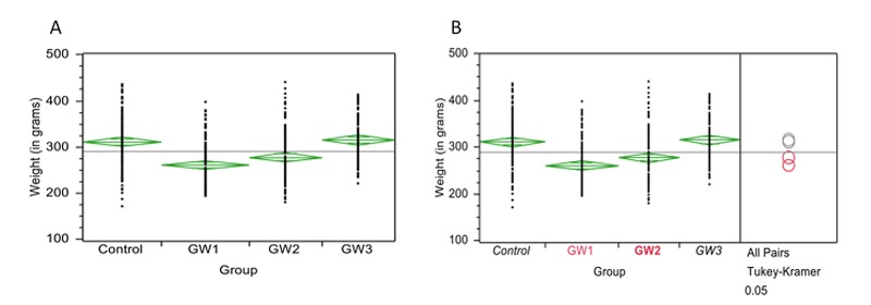
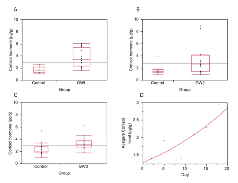
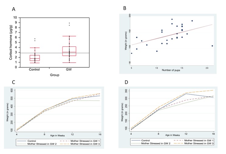
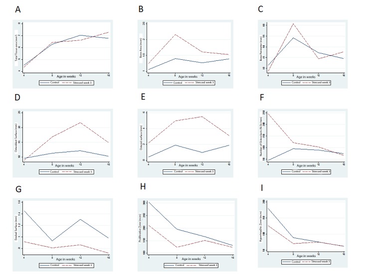
Unperturbed fetal development is essential for future health of an individual. Previous studies have linked diseases of aging to harmful alterations that happen during fetal development. Given the significant long-term impact that intrauterine environment has on an individual's life, it was hypothesized that maternal stress during pregnancy will have negative effects on the offspring's prenatal and postnatal growth. To test this, twenty-eight female and seven male Wistar rats (Rattus norvegicus) were purchased and bred to produce 176 offspring. During pregnancy, dams were randomly divided into four groups (n=7, per group) and immobilization stress induced as follows; Group 1 (GW1): immobilization stress on days 1-7 of pregnancy, Group 2 (GW2): on days 8-14, Group 3 (GW3): on days 15-21, Group 4 (Controls): left undisturbed. Maternal cortisol hormone, food intake, and weight gain were monitored during pregnancy. Pups were raised under normal laboratory conditions and sacrificed at ages: 4, 8, 12, and 16 weeks to determine the effect of prenatal stress. At necropsy, the tibia was removed and processed for histology. Differences among groups were determined by T-test or analysis of variance (ANOVA). Linear regression analysis was performed to establish the relationship between stress in utero and indicators of bone development in offspring. P values ≤ 0.05 were considered significant. Cortisol hormone levels in controls were lower than those of stressed animals. Stressed dams consumed 12.5% less food per day compared to controls. Animals in GW1 and GW2 gained less weight during pregnancy but had larger litters than did GW3 or the control group. Offspring born to GW3 were heavier compared to all other groups. GW3 offspring had a higher rate of bone formation. In conclusion, stress during pregnancy resulted in increased cortisol and reduced food intake in mothers, but faster growth and higher weight gain in offspring compared to controls.
Keywords: elevated cortisol; offspring development; prenatal stress.
Figures
References
-
- Barker DJ. Intrauterine programming of adult disease. Mol Med Today. 1995;1:418–423. - PubMed
-
- Dennison E, Fall C, Cooper C, Barker D. Prenatal factors influencing long-term outcome. Horm Res. 1997;48(Suppl 1):25–29. - PubMed
-
- Barker DJ. The fetal and infant origins of disease. Eur J Clin Invest. 1995;25:457–463. - PubMed
-
- Lazinski MJ, Shea AK, Steiner M. Effects of maternal prenatal stress on offspring development: a commentary. Arch Womens Ment Health. 2008;11:363–375. - PubMed
-
- Field T, Diego M. Cortisol: the culprit prenatal stress variable. Int J Neurosci. 2008;118:1181. - PubMed
LinkOut - more resources
Full Text Sources
Other Literature Sources