. 2014 Jan 6:4:77-89.
doi: 10.1016/j.fob.2014.01.002. eCollection 2014.

Ebselen induces reactive oxygen species (ROS)-mediated cytotoxicity in Saccharomyces cerevisiae with inhibition of glutamate dehydrogenase being a target

Affiliations

Ebselen induces reactive oxygen species (ROS)-mediated cytotoxicity in Saccharomyces cerevisiae with inhibition of glutamate dehydrogenase being a target

Gajendra Kumar Azad et al. FEBS Open Bio. .

Erratum in

Abstract

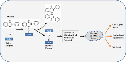
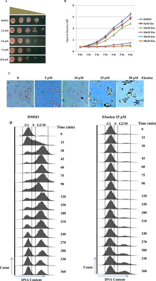
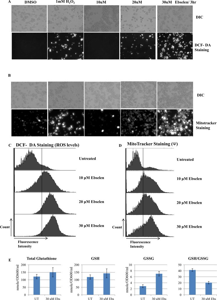
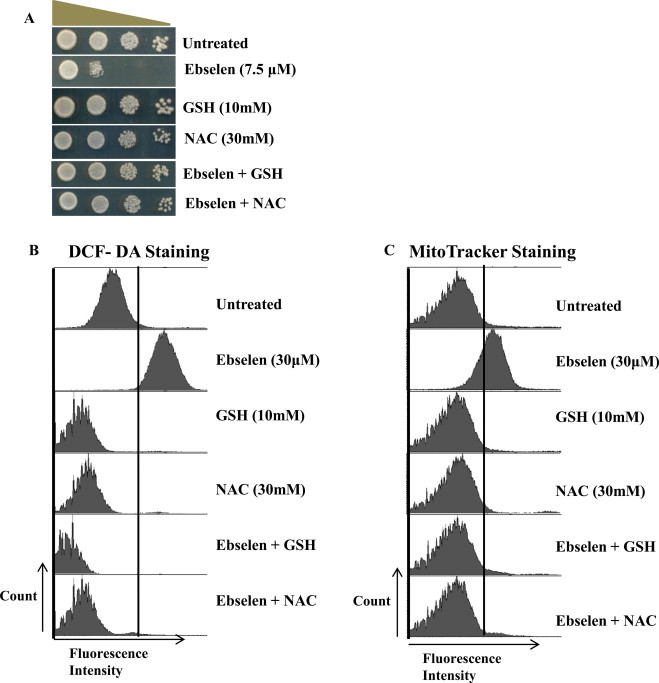
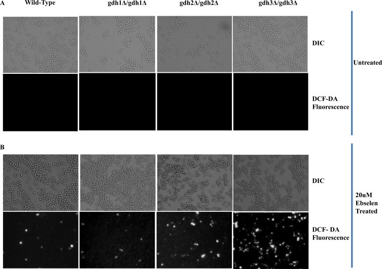
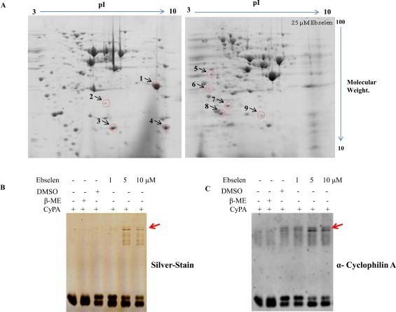
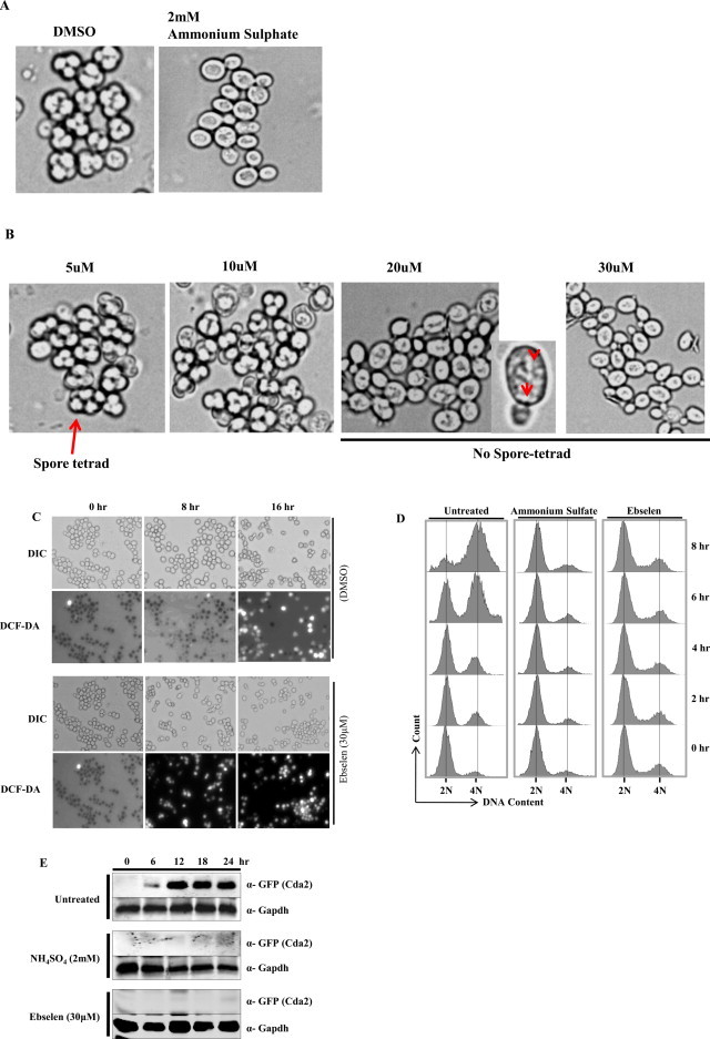
Ebselen is a synthetic, lipid-soluble seleno-organic compound. The high electrophilicity of ebselen enables it to react with multiple cysteine residues of various proteins. Despite extensive research on ebselen, its target molecules and mechanism of action remains less understood. We performed biochemical as well as in vivo experiments employing budding yeast as a model organism to understand the mode of action of ebselen. The growth curve analysis and FACS (florescence activated cell sorting) assays revealed that ebselen exerts growth inhibitory effects on yeast cells by causing a delay in cell cycle progression. We observed that ebselen exposure causes an increase in intracellular ROS levels and mitochondrial membrane potential, and that these effects were reversed by addition of antioxidants such as reduced glutathione (GSH) or N-acetyl-l-cysteine (NAC). Interestingly, a significant increase in ROS levels was noticed in gdh3-deleted cells compared to wild-type cells. Furthermore, we showed that ebselen inhibits GDH function by interacting with its cysteine residues, leading to the formation of inactive hexameric GDH. Two-dimensional gel electrophoresis revealed protein targets of ebselen including CPR1, the yeast homolog of Cyclophilin A. Additionally, ebselen treatment leads to the inhibition of yeast sporulation. These results indicate a novel direct connection between ebselen and redox homeostasis.

Keywords: CypA, Cyclophilin A; DCFH-DA, 2,7-dichlorodihydrofluorescein diacetate; Ebselen; FACS, florescence activated cell sorting; GDH, glutamate dehydrogenase; GSH, glutathione; Glutamate dehydrogenase; Histone clipping; Mitochondrial membrane potential; NAC, N-acetyl-l-cysteine; Ni-NTA, nickel-nitrilotriacetic acid; ROS levels; ROS, reactive oxygen species; SOD, superoxide dismutase; Yeast sporulation.


Figures

None
Graphical abstract
Fig. 1
Fig. 1
Increasing concentration of ebselen inhibits growth of yeast cells: (A) Spot test of wild type cells (WT1588-4C) in presence of DMSO (control), 2.5, 5.0, 7.5 and 10.0 μM ebselen. Yeast saturated cultures were serially diluted (10−1, 10−2, 10−3, 10−4) in 1.0 ml of sterile double distilled water. (B) Wild type yeast cells were grown in YPD medium until log phase reached at OD600 (0.6–0.8) then treated with ebselen at different concentrations (5, 10, 20, 30 and 50 μM) for 6 h. Growth was recorded by taking aliquots at regular interval. (C) Methylene blue assay was performed in treated and untreated cells and observed under microscope with magnification 400×. (D) FACS analysis showing the effect of the ebselen on yeast cell cycle progression. Wild-type cells were cultured in SC medium to exponential phase and treated with alpha factor to synchronize all cells in G1 phase. After synchronization cells were released in either DMSO (control) or 25 μM ebselen containing media. The culture was sampled at indicated time points and cellular DNA content was analyzed by FACS.
Fig. 2
Fig. 2
Ebselen treatment increases reactive oxygen species production and mitochondrial membrane potential in S. cerevisiae cells. ROS production detected by (A) DCF-DA and mitochondrial membrane potential by MitoTracker (B) in control cells and cells treated for 3 h. Cells were treated with 1 mM H2O2 for 3 h and it served as positive control. In A and B the upper panels show phase contrast microscopy; the lower panels show fluorescence microscopy of the same cells. (C and D) Yeast wild type strain was grown in SC media till exponential phase. Cells were treated with indicated concentration of ebselen for 3 h. The cells were then stained with DCF-DA or MitoTracker Red and examined by FACS as described in materials and methods. (E) Wild type cells were grown in DMSO or indicated concentration of ebselen for 3 h. GSH, GSSG, and the GSH:GSSG ratios were determined. Values are means S.D. of three independent cultures.
Fig. 3
Fig. 3
Effect of exogenous supply of GSH or NAC on ebselen induced ROS accumulation. (A) Spot test of wild type cells (WT1588-4C) supplemented with either NAC or GSH, and plates containing ebselen (7.5 μM) with either NAC or GSH. Yeast saturated cultures were serially diluted (10−1, 10−2, 10−3, 10−4) in 1.0 ml of sterile double distilled water and spotted onto the plates. Cells were cultured at 30 °C for 2–3 days. (D and E) Wild-type yeast strain grown in SC media supplemented with or without 10 mM GSH or 20 mM NAC for 1 h followed by exposure to 30 μM ebselen for 3 h. Yeast cells were processed for FACS analysis after staining with either DCF-DA (D) or MitoTracker Red (E).
Fig. 4
Fig. 4
gdh3-dependent ROS generation by ebselen in S. cerevisiae. Wild-type and the gdh1, gdh2 and gdh3-deletion mutant were treated with 20 μM ebselen for 2 h. Then, they were stained with 10 μM H2DCFDA for 1 min, and the level of ROS was observed by fluorescence microscopy. (A) Background ROS levels in WT, gdh1, gdh2 and gdh3-deletion mutants. Upper panels show phase contrast microscopy; the lower panels show florescence microscopy of the same cells. (B) ROS level in mutant and wild-type cells.
Fig. 5
Fig. 5
The effect of ebselen on GDH. (A) The effect of ebselen on GDH protease activity was analyzed by incubating purified core histones with chicken GDH in presence and/or absence of β-mercaptoethanol. (B and C) Effect of ebselen on core histones and on GDH protein profile was analyzed by resolving proteins on non-reducing and reducing SDS–PAGE, followed by coomassie brilliant blue R staining (B) and by western blotting with anti-GDH antibody (C) respectively. (D) The cysteine residues (labeled in red) of GDH were highlighted as the probable interacting site for ebselen. (For interpretation of the references to color in this figure legend, the reader is referred to the web version of this article.).
Fig. 6
Fig. 6
Ebselen alters the proteome profile of yeast cells: two dimensional gel electrophoresis gels are showing differentially expressed proteins in control (DMSO) and 25 μM ebselen treatment. (A) Proteins were separated in the first dimension on IEF gel (7 cm, pH 3–10) and then run on 12% SDS–PAGE. The red circles represent the protein spots which were excised from the gel for mass spectrometric analysis. The effect of ebselen on recombinant human Cyclophilin A. 5.0 μg CyPA incubated with increasing concentration of ebselen for 30 min, followed by running non-reducing SDS–PAGE (B) silver stained photograph, (C) corresponding cyclophilin A western signal image. Arrows indicate cyclophilin A complex. (For interpretation of the references to color in this figure legend, the reader is referred to the web version of this article.).
Fig. 7
Fig. 7
Ebselen strongly inhibits sporulation in yeast. (A) Microscopic images of cells sporulated for 24 h in the absence of drug (control), in the presence of 2 mM ammonium sulfate, (B) or increasing concentration of ebselen. Part of the 20 μM ebselen treated image was magnified; arrows indicate granular bodies of unknown origin. (C) Microscopic images of cells sporulated for indicated time (0, 8 and 16 h) in the absence of drug (DMSO), in the presence of 30 μM ebselen. The upper panels show phase contrast microscopy; the lower panels show fluorescence microscopy of the same cells after staining with DCF-DA. (D) Analysis of pre-meiotic DNA synthesis in a control (DMSO), and cells treated with ammonium sulfate (2 mM) or Ebselen (30 μM) through FACS. Samples were taken at regular interval as indicated in figure after induction of sporulation. Samples were subjected to FACS analysis and results were processed with BD FACS Diva software. (E) Yeast strain USY613 (USY61+ pCDA2-eGFP::HygB) was cultured as described in materials and methods and treated with 30 μM of ebselen or 2 mM ammonium sulfate for 24 h. 10 ml cells were harvested at regular intervals (0, 6, 12, 18, 24 h). Whole cell extracts were prepared by TCA extraction method and samples were subjected to western blot anlaysis using indicated antibodies. Tbp and Gapdh served as loading controls.

References

    1. Aon M.A., Cortassa S., O’Rourke B. Redox-optimized ROS balance: a unifying hypothesis. Biochim. Biophys. Acta. 2010;1797:865–877. - PMC - PubMed
    1. Hu J., Dong L., Outten C.E. The redox environment in the mitochondrial intermembrane space is maintained separately from the cytosol and matrix. J. Biol. Chem. 2008;283:29126–29134. - PMC - PubMed
    1. Go Y.M., Jones D.P. Redox compartmentalization in eukaryotic cells. Biochim. Biophys. Acta. 2008;1780:1273–1290. - PMC - PubMed
    1. Cecarini V., Gee J., Fioretti E., Amici M., Angeletti M., Eleuteri A.M., Keller J.N. Protein oxidation and cellular homeostasis: emphasis on metabolism. Biochim. Biophys. Acta. 2007;1773:93–104. - PubMed
    1. Avery S.V. Molecular targets of oxidative stress. Biochem. J. 2011;434:201–210. - PubMed