. 2014 Feb 4;16(1):R41.
doi: 10.1186/ar4470.

Generation of disease-specific induced pluripotent stem cells from patients with rheumatoid arthritis and osteoarthritis

Generation of disease-specific induced pluripotent stem cells from patients with rheumatoid arthritis and osteoarthritis

Jaecheol Lee et al. Arthritis Res Ther. .

Abstract

Introduction: Since the concept of reprogramming mature somatic cells to generate induced pluripotent stem cells (iPSCs) was demonstrated in 2006, iPSCs have become a potential substitute for embryonic stem cells (ESCs) given their pluripotency and "stemness" characteristics, which resemble those of ESCs. We investigated to reprogram fibroblast-like synoviocytes (FLSs) from patients with rheumatoid arthritis (RA) and osteoarthritis (OA) to generate iPSCs using a 4-in-1 lentiviral vector system.

Methods: A 4-in-1 lentiviral vector containing Oct4, Sox2, Klf4, and c-Myc was transduced into RA and OA FLSs isolated from the synovia of two RA patients and two OA patients. Immunohistochemical staining and real-time PCR studies were performed to demonstrate the pluripotency of iPSCs. Chromosomal abnormalities were determined based on the karyotype. SCID-beige mice were injected with iPSCs and sacrificed to test for teratoma formation.

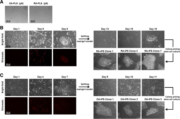
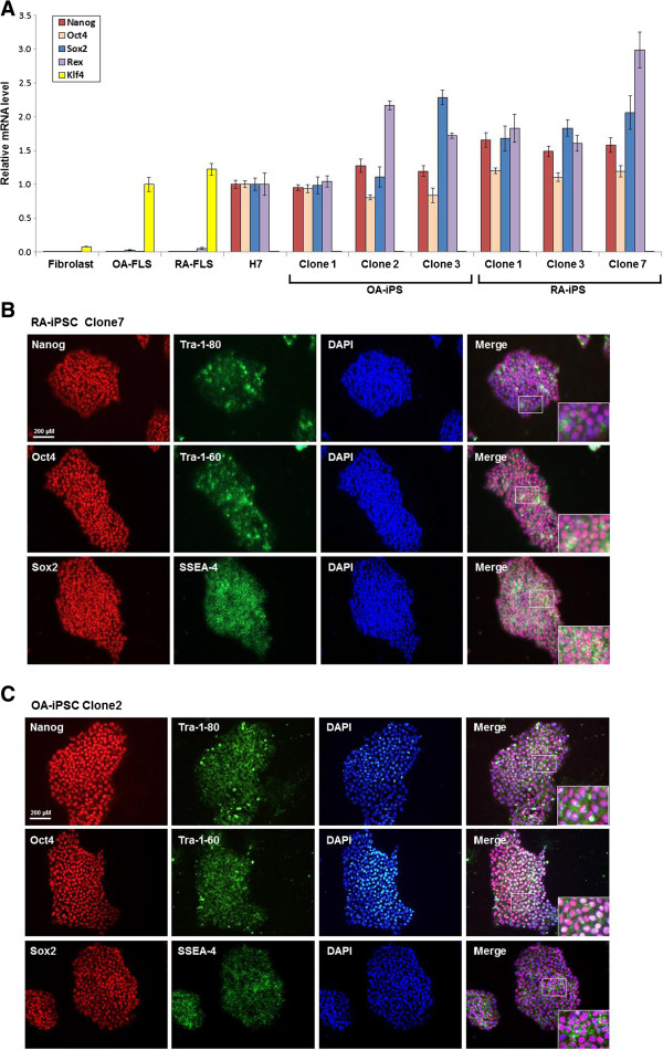
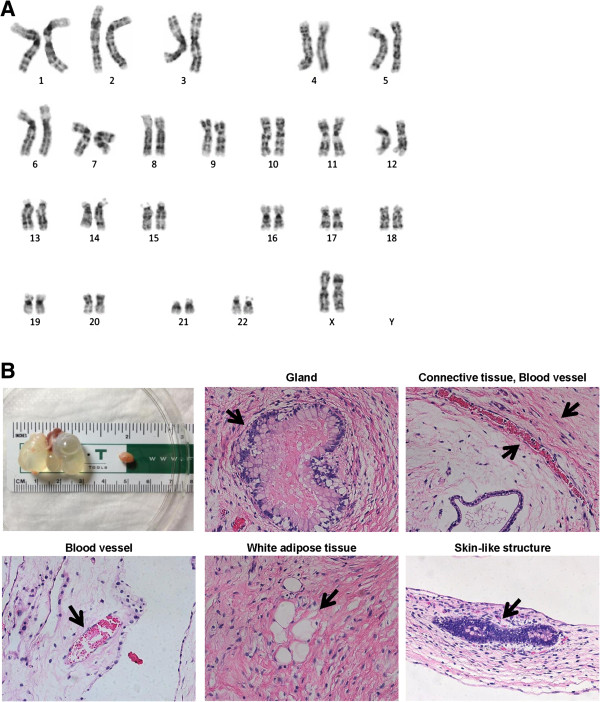
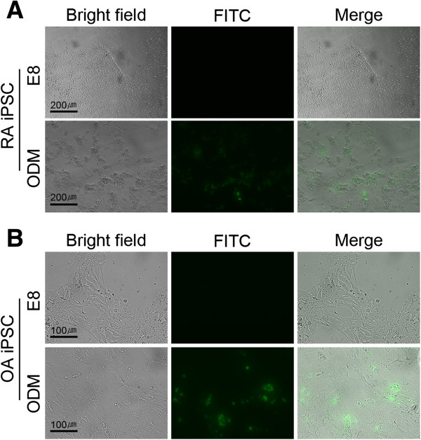
Results: After 14 days of transduction using the 4-in-1 lentiviral vector, RA FLSs and OA FLSs were transformed into spherical shapes that resembled embryonic stem cell colonies. Colonies were picked and cultivated on matrigel plates to produce iPSC lines. Real-time PCR of RA and OA iPSCs detected positive markers of pluripotency. Immunohistochemical staining tests with Nanog, Oct4, Sox2, Tra-1-80, Tra-1-60, and SSEA-4 were also positive. Teratomas that comprised three compartments of ectoderm, mesoderm, and endoderm were formed at the injection sites of iPSCs. Established iPSCs were shown to be compatible by karyotyping. Finally, we confirmed that the patient-derived iPSCs were able to differentiate into osteoblast, which was shown by an osteoimage mineralization assay.

Conclusion: FLSs derived from RA and OA could be cell resources for iPSC reprogramming. Disease- and patient-specific iPSCs have the potential to be applied in clinical settings as source materials for molecular diagnosis and regenerative therapy.


Figures

Figure 1
Figure 1
Successful generation of induced pluripotent stem cells from rheumatoid arthritis and osteoarthritis fibroblast-like synoviocytes. (A) Rheumatoid arthritis (RA) fibroblast-like synoviocytes (FLSs) and osteoarthritis (OA) FLSs were cultured before reprogramming. RA and OA FLS was extracted from RA patients (n = 2) and OA patients (n = 2) and were cultured as previously documented. There were five passages of RA and OA FLSs. Synovium was gathered from the elbow and knee during synovectomy surgery. One sample (RA1) was from a disease-modifying antirheumatic drug-naïve patient and the other sample (RA2) from a biologics-refractory patient with persistent high disease activity. (B) Tomato fluorescence was shown shortly after virus infection. On the 6th day after the transduction of four-in-one viral vector on RA FLSs, colony-like spheres began to form. On the 18th day, colonies were picked and resuspended on Matrigel-coated culture dishes. (C) Tomato fluorescence was positively shown on four-in-one vector-transduced OA FLSs. On the 7th day after the transduction of four-in-one viral vector on OA FLS, colony-like spheres began to form. On the 11th day, colonies were picked and resuspended on Matrigel-coated culture dishes.
Figure 2
Figure 2
Positive expression of pluripotency marker on osteoarthritis and rheumatoid arthritis induced pluripotent stem cells. (A) The expression of stemness genes in osteoarthritis (OA) induced pluripotent stem cells (iPSCs) and rheumatoid arthritis (RA) iPSCs was analyzed by quantitative reverse transcriptase polymerase chain reaction (RT-PCR) analysis. Total RNA of fibroblast, OA fibroblast-like synoviocytes (FLSs), RA FLSs, H7, OA iPSCs and RA iPSCs was isolated and analyzed for endogenous pluripotent genes by RT-PCR analysis. The expression of endogenous Nanog, Oct4, Sox2 and Rex is upregulated in OA iPSCs and RA iPSCs. OA FLS and RA FLS already expressed Klf4 before reprogramming process started. (B) Immunofluorescence staining against Nanog, Oct4, Sox2, Tra-1-80, Tra-1-60, and SSEA-4 in RA iPSCs. RA iPSCs expressed a high level of these markers. (C) Immunofluorescence staining against Nanog, Oct4, Sox2, Tra-1-80, Tra-1-60, and SSEA-4 in OA iPSCs. OA iPSCs were positive for the pluripotency genes such as Nanog, Oct4, Sox2, Tra-1-80, Tra-1-60, and SSEA-4.
Figure 3
Figure 3
Characterization of rheumatoid arthritis induced pluripotent stem cells. (A) High-resolution, G-banded karyotype indicated a normal diploid female chromosomal content 46XX in rheumatoid arthritis (RA) induced pluripotent stem cells (iPSCs). (B) Teratoma formation occurred after injection of RA iPSCs into immunocompromised mice. Tumors were detected from the sites of injection and harvested after 3 months, and were examined for the presence of cells of three embryonic germ layers. Teratomas contained tissue from all three germ layers, including the gland (endoderm), blood vessel (mesoderm), adipose tissue (mesoderm), and skin-like structure (ectoderm).
Figure 4
Figure 4
In vitro osteogenic differentiation of rheumatoid arthritis and osteoarthritis induced pluripotent stem cells. To induce osteogenic differentiation, rheumatoid arthritis (RA) and osteoarthritis (OA) induced pluripotent stem cells (iPSCs) were cultured in E8 medium or osteogenic differentiation medium (ODM) for 7 days. Osteogenic differentiation was determined by the fluorescence detection for mineralization. Osteogenic mineralization was detected in both ODM-treated RA iPSCs (A) and OA iPSCs (B) by fluorescence microscope (fluorescein isothiocyanate (FITC)).

Similar articles

Cited by

References

    1. Takahashi K, Yamanaka S. Induction of pluripotent stem cells from mouse embryonic and adult fibroblast cultures by defined factors. Cell. 2006;16:663–676. doi: 10.1016/j.cell.2006.07.024. - DOI - PubMed
    1. Puri MC, Nagy A. Concise review: Embryonic stem cells versus induced pluripotent stem cells: the game is on. Stem Cells. 2012;16:10–14. doi: 10.1002/stem.788. - DOI - PubMed
    1. Bilic J, Izpisua Belmonte JC. Concise review: Induced pluripotent stem cells versus embryonic stem cells: close enough or yet too far apart? Stem Cells. 2012;16:33–41. doi: 10.1002/stem.700. - DOI - PubMed
    1. Tiscornia G, Vivas EL, Izpisúa Belmonte JC. Diseases in a dish: modeling human genetic disorders using induced pluripotent cells. Nat Med. 2011;16:1570–1576. - PubMed
    1. Grskovic M, Javaherian A, Strulovici B, Daley GQ. Induced pluripotent stem cells – opportunities for disease modelling and drug discovery. Nat Rev Drug Discov. 2011;16:915–929. - PubMed

Publication types