Case Reports
. 2014 Feb 3:14:13.
doi: 10.1186/1471-2415-14-13.

Case report: an atypical peripapillary uveal melanoma

Affiliations
Case Reports

Case report: an atypical peripapillary uveal melanoma

Li-Anne Lim et al. BMC Ophthalmol. .

Abstract

Background: The treatment of uveal melanoma has seen a shift towards eye conserving treatments. Efforts have been made towards the identification of patients at high risk of metastatic disease with the use of prognostic fine needle biopsy, Monosomy 3 a risk factor for metastatic death thought to occur early in the development of uveal melanoma.

Case presentation: We report a case in which an atypical optic nerve lesion was found to be a peripapillary primary uveal melanoma with distinct non-pigmented and pigmented halves on gross dissection and corresponding disomy 3 and monosomy 3 halves. The tumour demonstrated rapid growth with apparent transformation from disomy 3 to monosomy 3.

Conclusions: These are clinical features that challenge the current concepts of the cytogenetic pathogenesis of uveal melanoma and demonstrate the potential problems and limitations of prognostic fine needle biopsy and molecular classifications.


Figures
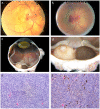
Figure 1
Figure 1
Clinical and pathological findings. (a) Initial dilated fundus examination: disc edema, hemorrhage and a serous retinal detachment. (b) Fundus examination 10 months later: increase in the size of the mass, with disc border hemorrhage and an adjacent ring of pigmentation. (c) (d) Gross pathology: uveal melanoma with distinct non-pigmented and pigmented halves. (e) Histopathological section of the non-pigmented half of the uveal melanoma, spindle cell type. (f) Histopathological section of the pigmented half of the uveal melanoma, of spindle cell type with melanin.
Figure 2
Figure 2
Imaging and immunohistochemical findings. (a) T1 weighted MRI of the globe: an elevated intraocular lesion at the posterior pole of moderately high signal. (b) B-scan ultrasound: dome shaped lesion at the posterior pole. (c) A-scan ultrasound: solid lesion at the posterior pole with low internal reflectivity and a kappa angle. (d) Immunohistochemistry: HMB-45 positive staining.

References

    1. Prescher G, Bornfeld N, Hirche H. et al.Prognostic implications of monosomy 3 in uveal melanoma. Lancet. 1996;347(9010):1222–1225. doi: 10.1016/S0140-6736(96)90736-9. - DOI - PubMed
    1. Prescher G, Bornfeld N, Becher R. Two subclones in a case of uveal melanoma. Relevance of monosomy 3 and multiplication of chromosome 8q. Canc Genet Cytogenet. 1994;77(2):144–146. doi: 10.1016/0165-4608(94)90230-5. - DOI - PubMed
    1. Wiltshire RN, Elner VM, Dennis T, Vine AK, Trent JM. Cytogenetic analysis of posterior uveal melanoma. Canc Genet Cytogenet. 1993;66(1):47–53. doi: 10.1016/0165-4608(93)90148-F. - DOI - PubMed
    1. Tschentscher F, Husing J, Holter T. et al.Tumor classification based on gene expression profiling shows that uveal melanomas with and without monosomy 3 represent two distinct entities. Canc Res. 2003;63(10):2578–2584. - PubMed
    1. Onken MD, Worley LA, Harbour JW. A metastasis modifier locus on human chromosome 8p in uveal melanoma identified by integrative genomic analysis. Clin Canc Res. 2008;14(12):3737–3745. doi: 10.1158/1078-0432.CCR-07-5144. - DOI - PubMed

Publication types

Supplementary concepts