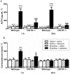
Corticotropin-releasing factor (CRF) receptor-1 is involved in cardiac noradrenergic activity observed during naloxone-precipitated morphine withdrawal
- PMID: 24490859
- PMCID: PMC3969081
- DOI: 10.1111/bph.12511
Corticotropin-releasing factor (CRF) receptor-1 is involved in cardiac noradrenergic activity observed during naloxone-precipitated morphine withdrawal
Abstract
Background and purpose: The negative affective states of withdrawal involve the recruitment of brain and peripheral stress circuitry [noradrenergic activity, induction of the hypothalamic-pituitary-adrenocortical (HPA) axis and activation of heat shock proteins (Hsps)]. Corticotropin-releasing factor (CRF) pathways are important mediators in the negative symptoms of opioid withdrawal. We performed a series of experiments to characterize the role of the CRF₁ receptor in the response of stress systems to morphine withdrawal and its effect in the heart using genetically engineered mice lacking functional CRF₁ receptors.
Experimental approach: Wild-type and CRF₁ receptor-knockout mice were treated with increasing doses of morphine. Precipitated withdrawal was induced by naloxone. Plasma adrenocorticotropic hormone (ACTH) and corticosterone levels, the expression of myocardial Hsp27, Hsp27 phosphorylated at Ser⁸², membrane (MB)- COMT, soluble (S)-COMT protein and NA turnover were evaluated by RIA, immunoblotting and HPLC.
Key results: During morphine withdrawal we observed an enhancement of NA turnover in parallel with an increase in mean arterial blood pressure (MAP) and heart rate (HR) in wild-type mice. In addition, naloxone-precipitated morphine withdrawal induced an activation of HPA axis and Hsp27. The principal finding of the present study was that plasma ACTH and corticosterone levels, MB-COMT, S-COMT, NA turnover, and Hsp27 expression and activation observed during morphine withdrawal were significantly inhibited in the CRF₁ receptor-knockout mice.
Conclusion and implications: Our results demonstrate that CRF/CRF₁ receptor activation may contribute to stress-induced cardiovascular dysfunction after naloxone-precipitated morphine withdrawal and suggest that CRF/CRF₁ receptor pathways could contribute to cardiovascular disease associated with opioid addiction.
Keywords: CRF1 receptor knockout mice; Hsp 27; NA turnover; heart; naloxone-precipitated morphine withdrawal.
© 2013 The British Pharmacological Society.
Figures


Similar articles
-
Cardiac Protective Role of Heat Shock Protein 27 in the Stress Induced by Drugs of Abuse.Int J Mol Sci. 2020 May 21;21(10):3623. doi: 10.3390/ijms21103623. Int J Mol Sci. 2020. PMID: 32455528 Free PMC article. Review.
-
Morphine withdrawal activates hypothalamic-pituitary-adrenal axis and heat shock protein 27 in the left ventricle: the role of extracellular signal-regulated kinase.J Pharmacol Exp Ther. 2012 Sep;342(3):665-75. doi: 10.1124/jpet.112.193581. Epub 2012 May 30. J Pharmacol Exp Ther. 2012. PMID: 22647273
-
Cardiac adverse effects of naloxone-precipitated morphine withdrawal on right ventricle: role of corticotropin-releasing factor (CRF) 1 receptor.Toxicol Appl Pharmacol. 2014 Feb 15;275(1):28-35. doi: 10.1016/j.taap.2013.12.021. Epub 2014 Jan 5. Toxicol Appl Pharmacol. 2014. PMID: 24398105
-
CRF₂ mediates the increased noradrenergic activity in the hypothalamic paraventricular nucleus and the negative state of morphine withdrawal in rats.Br J Pharmacol. 2011 Feb;162(4):851-62. doi: 10.1111/j.1476-5381.2010.01090.x. Br J Pharmacol. 2011. PMID: 20973778 Free PMC article.
-
Corticotropin releasing factor (CRF) receptor signaling in the central nervous system: new molecular targets.CNS Neurol Disord Drug Targets. 2006 Aug;5(4):453-79. doi: 10.2174/187152706777950684. CNS Neurol Disord Drug Targets. 2006. PMID: 16918397 Free PMC article. Review.
Cited by
-
Cardiac Protective Role of Heat Shock Protein 27 in the Stress Induced by Drugs of Abuse.Int J Mol Sci. 2020 May 21;21(10):3623. doi: 10.3390/ijms21103623. Int J Mol Sci. 2020. PMID: 32455528 Free PMC article. Review.
-
Corticotropin releasing factor excites neurons of posterior hypothalamic nucleus to produce tachycardia in rats.Sci Rep. 2016 Feb 1;6:20206. doi: 10.1038/srep20206. Sci Rep. 2016. PMID: 26831220 Free PMC article.
-
Binge Ethanol and MDMA Combination Exacerbates Toxic Cardiac Effects by Inducing Cellular Stress.PLoS One. 2015 Oct 28;10(10):e0141502. doi: 10.1371/journal.pone.0141502. eCollection 2015. PLoS One. 2015. PMID: 26509576 Free PMC article.
-
Sex differences between CRF1 receptor deficient mice following naloxone-precipitated morphine withdrawal in a conditioned place aversion paradigm: implication of HPA axis.PLoS One. 2015 Apr 1;10(4):e0121125. doi: 10.1371/journal.pone.0121125. eCollection 2015. PLoS One. 2015. PMID: 25830629 Free PMC article.
References
-
- Almela P, Martínez-Laorden E, Atucha NM, Milanés MV, Laorden ML. Naloxone-precipitated morphine withdrawal evokes phosphorylation of heat shock protein 27 in rat heart through extracellular signal-regulated kinase. J Mol Cell Cardiol. 2011;51:129–139. - PubMed
-
- Aquaro GD, Gabutti A, Meini M, Prontera C, Pasanisi E, Passino C, et al. Silent myocardial damage in cocaine addicts. Heart. 2011;97:2056–2062. - PubMed
-
- Arlt J, Jahn H, Kellner M, Strohle A, Yassouridis A, Wiedemann K. Modulation of sympathetic activity by corticotropin-releasing hormone and atria natriuretic peptide. Endocrinology. 2003;37:362–368. - PubMed
Publication types
MeSH terms
Substances
LinkOut - more resources
Full Text Sources
Other Literature Sources
Molecular Biology Databases
Research Materials
Miscellaneous

