. 2014 Feb 3;21(1):10.
doi: 10.1186/1423-0127-21-10.

Nitrogen-containing bisphosphonates inhibit RANKL- and M-CSF-induced osteoclast formation through the inhibition of ERK1/2 and Akt activation

Affiliations

Nitrogen-containing bisphosphonates inhibit RANKL- and M-CSF-induced osteoclast formation through the inhibition of ERK1/2 and Akt activation

Masanobu Tsubaki et al. J Biomed Sci. .

Abstract

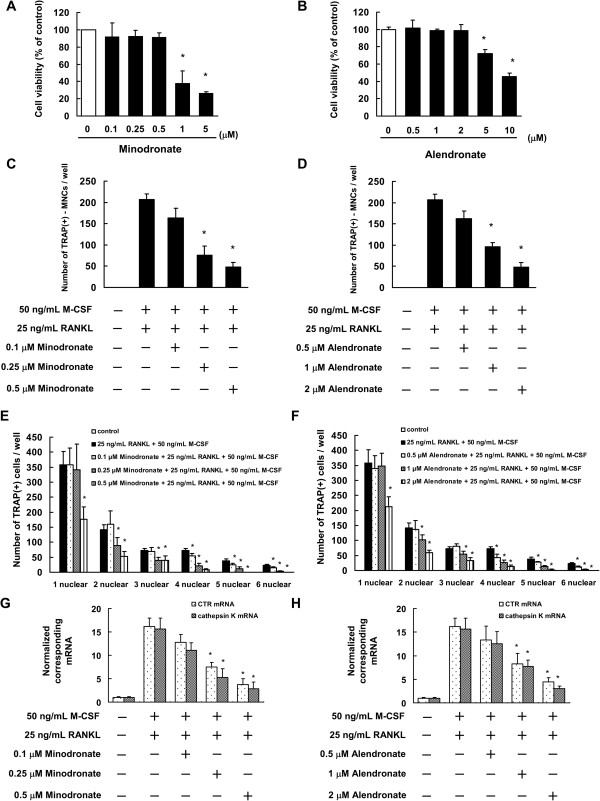
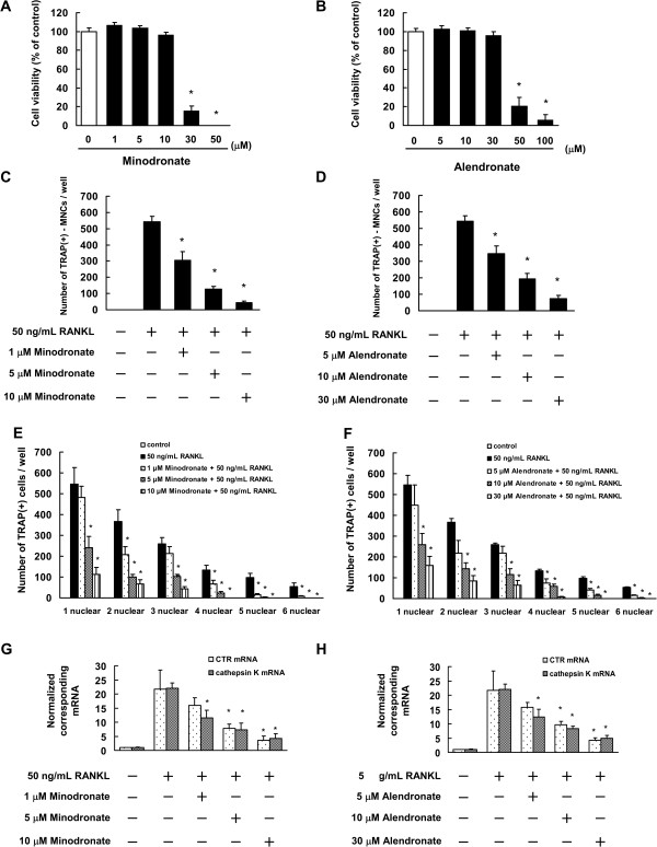
Background: Bisphosphonates are an important class of antiresorptive drugs used in the treatment of metabolic bone diseases. Recent studies have shown that nitrogen-containing bisphosphonates induced apoptosis in rabbit osteoclasts and prevented prenylated small GTPase. However, whether bisphosphonates inhibit osteoclast formation has not been determined. In the present study, we investigated the inhibitory effect of minodronate and alendronate on the osteoclast formation and clarified the mechanism involved in a mouse macrophage-like cell lines C7 and RAW264.7.

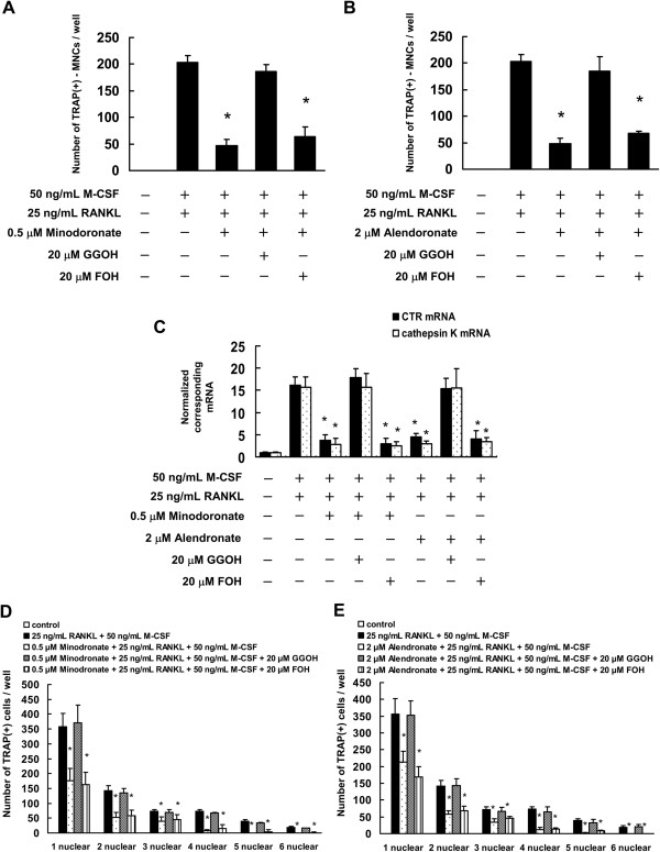
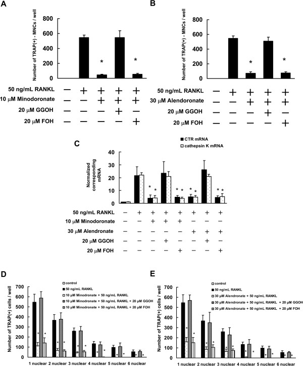
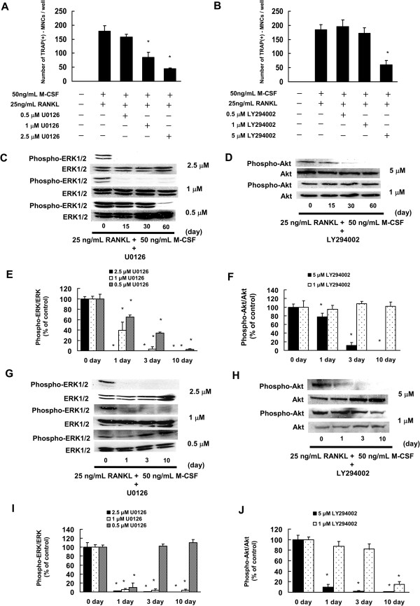
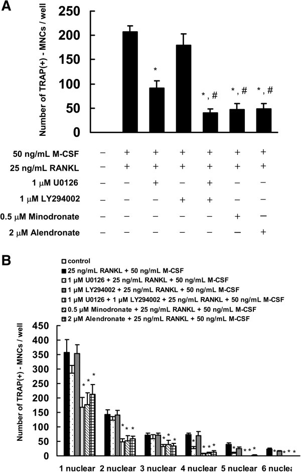
Results: It was found that minodronate and alendronate inhibited the osteoclast formation of C7 cells induced by receptor activator of NF-κB ligand and macrophage colony stimulating factor, which are inhibited by the suppression of geranylgeranyl pyrophosphate (GGPP) biosynthesis. It was also found that minodronate and alendronate inhibited the osteoclast formation of RAW264.7 cells induced by receptor activator of NF-κB ligand. Furthermore, minodronate and alendornate decreased phosphorylated extracellular signal-regulated kinase 1/2 (ERK1/2) and Akt; similarly, U0126, a mitogen protein kinase kinase 1/2 (MEK1/2) inhibitor, and LY294002, a phosphatidylinositol 3-kinase (PI3K) inhibitor, inhibited osteoclast formation.

Conclusions: This indicates that minodronate and alendronate inhibit GGPP biosynthesis in the mevalonate pathway and then signal transduction in the MEK/ERK and PI3K/Akt pathways, thereby inhibiting osteoclast formation. These results suggest a novel effect of bisphosphonates that could be effective in the treatment of bone metabolic diseases, such as osteoporosis.


Figures

Figure 1
Figure 1
Minodronate and alendronate inhibited osteoclast formation in C7 cells. (A, B) Determination of the appropriate concentrations of minodronate (A) and alendronate (B) that are not cytotoxic to C7 cells. Cells (5000 cells/well) were incubated in 96-well plates for 24 h and then treated with various concentrations of minodronate and alendronate. After 12 days, cell viability was quantified by conducting WST-8 assays. The results are representative of 5 independent experiments. *P < 0.01 compared to the controls. (C-F) Inhibition of osteoclast formation by minodronate and alendronate. C7 cells were cultured for 12 days and then treated with 0.1, 0.25, or 0.5 μM minodronate (C, E) or 0.5, 1, or 2 μM alendronate (D, F). Cells were cultured in the presence of 25 ng/mL RANKL plus 50 ng/mL M-CSF. Cultures were fed every 3 days by replacing with 500 μL of fresh medium with or without minodronate, alendronate, RANKL, and M-CSF. Cultures were fixed and stained for TRAP-positive multinucleated cells (C, D), and TRAP-positive cells (E, F) per well was counted. These results are representative of 5 independent experiments. *P < 0.01 compared to 25 ng/mL RANKL plus 50 ng/mL M-CSF administration. (G, H) Inhibitory effect of minodronate and alendronate on RANKL and M-CSF-induced CTR and cathepsin K mRNA expression. C7 cells were treated with minodronate (G) or alendronate (H) with 25 ng/mL RANKL plus 50 ng/mL M-CSF for 12 days. Total RNA was extracted and the levels of CTR and cathepsin K mRNA expression were determined by real-time PCR. The results are expressed as the ratio of treated to control samples after normalization to GAPDH mRNA levels. The results are representative of 4 independent experiments. *P < 0.01 compared to 25 ng/mL RANKL plus 50 ng/mL M-CSF administration.
Figure 2
Figure 2
Minodronate and alendronate inhibited osteoclast formation in RAW264.7 cells. (A, B) Determination of the appropriate concentrations of minodronate (A) and alendronate (B) that are not cytotoxic to RAW264.7 cells. Cells (5000 cells/well) were incubated in 96-well plates for 24 h and then treated with various concentrations of minodronate and alendronate. After 7 days, cell viability was quantified by conducting WST-8 assays. The results are representative of 4 independent experiments. *P < 0.01 compared to the controls. (C-F) Inhibition of osteoclast formation by minodronate and alendronate. RAW264.7 cells were cultured for 12 days and then treated with 1, 5, or 10 μM minodronate (C, E) or 5, 10, or 20 μM alendronate (D, F). Cells were cultured in the presence of 50 ng/mL RANKL. Cultures were fed every 2 days by replacing with 500 μL of fresh medium, with or without minodronate, alendronate, and RANKL. Cultures were fixed and stained for TRAP-positive multinucleated cells (C, D), and TRAP-positive cells (E, F) per well was counted. These results are representative of 4 independent experiments. *P < 0.01 compared to 50 ng/mL RANKL administration. (G, H) Inhibitory effect of minodronate and alendronate on RANKL and M-CSF-induced CTR and cathepsin K mRNA expression. RAW264.7 cells were treated with minodronate (G) and alendronate (H) with 50 ng/mL RANKL for 7 days. Total RNA was extracted, and the CTR and cathepsin K mRNA levels were determined by real-time PCR. The results are expressed as the ratio of treated to control samples after normalization to GAPDH mRNA levels. The results are representative of 4 independent experiments. *P < 0.01 compared to 50 ng/mL RANKL administration.
Figure 3
Figure 3
Minodronate and alendronate inhibited osteoclast formation via suppression of GGPP biosynthesis in C7 cells. C7 cells were pretreated with 20 μM FOH or 20 μM GGOH for 4 h and were then treated with 0.5 μM minodronate or 2 μM alendronate and 25 ng/mL RANKL plus 50 ng/mL M-CSF for 12 days. Cultures were fed every 3 days by replacing with 500 μL of fresh medium, with or without minodronate, alendronate, RANKL, M-CSF, FOH, and GGOH. (A, B) Inhibitory effect of mevalonate pathway intermediates FOH or GGOH on the inhibition of osteoclast formation by minodronate (A) and alendronate (B). Cultures were fixed and stained for TRAP-positive multinucleated cells, and the number of cells per well was counted. These results are representative of 5 independent experiments. *P < 0.01 compared to 25 ng/mL RANKL plus 50 ng/mL M-CSF administration. (C) CTR and cathepisin K mRNA expression in C7 cells that were treated with minodronate or alendronate along with FOH or GGOH. Total RNA was extracted, and the CTR and cathepsin K mRNA levels were determined by real-time PCR. The results are expressed as the ratio of treated to control samples after normalization to GAPDH mRNA levels. The results are representative of 4 independent experiments. *P < 0.01 compared to 25 ng/mL RANKL plus 50 ng/mL M-CSF administration. (D, E) Cultures were fixed and stained for TRAP-positive cells, and the number of cells per well was counted. These results are representative of 4 independent experiments. *P < 0.01 compared to 25 ng/mL RANKL plus 50 ng/mL M-CSF administration.
Figure 4
Figure 4
Minodronate and alendronate inhibited osteoclast formation via suppression of GGPP biosynthesis in RAW264.7 cells. RAW264.7 cells were pretreated with 20 μM FOH or 20 μM GGOH for 4 h and then treated with 10 μM minodronate or 30 μM alendronate, and 50 ng/mL RANKL for 7 days. Cultures were fed every 2 days by replacing with 500 μL of fresh medium, with or without minodronate, alendronate, RANKL, FOH, and GGOH. (A, B) Inhibitory effect of mevalonate pathway intermediates FOH or GGOH on the inhibition of osteoclast formation by minodronate (A) or alendronate (B). Cultures were fixed and stained for TRAP-positive multinucleated cells, and the number of cells per well was counted. These results are representative of 5 independent experiments. *P < 0.01 compared to 50 ng/mL RANKL administration. (C) CTR and cathepsin K mRNA expression in RAW264.7 cells that were treated with minodronate or alendronate along with FOH or GGOH. Total RNA was extracted, and the CTR and cathepsin K mRNA levels were determined by real-time PCR. The results are expressed as the ratio of treated to control samples after normalization to GAPDH mRNA levels. The results are representative of 4 independent experiments. *P < 0.01 compared to 50 ng/mL RANKL administration. (D, E) Cultures were fixed and stained for TRAP-positive cells, and the number of cells per well was counted. These results are representative of 4 independent experiments. *P < 0.01 compared to 50 ng/mL RANKL administration.
Figure 5
Figure 5
RANKL plus M-CSF induced activation of ERK1/2 and Akt in C7 cells, which could be inhibited by minodronate and alendronate. (A, B) C7 cells were cultured in the presence of 25 ng/mL RANKL plus 50 ng/mL M-CSF for 15, 30, and 60 min (A) or 1, 3, and 10 days (B). (C, D) C7 cells were treated with 0.5 μM minodronate for 24 h. Cells were cultured in the presence of 25 ng/mL RANKL plus 50 ng/mL M-CSF for 15, 30, and 60 min (C) or 1, 3, and 10 days (D). (E, F) C7 cells were treated with 2 μM alendronate for 24 h. Cells were cultured in the presence of 25 ng/mL RANKL plus 50 ng/mL M-CSF for 15, 30, and 60 min (E) or 1, 3, and 10 days (F). Whole cell lysates were generated and immunoblotted with an antibody against phosphorylated ERK1/2 (phospho-ERK1/2), phosphorylated Akt (phospho-Akt), phosphorylated p38MAPK (phospho-p38MAPK), ERK1/2, Akt, and p38MAPK. (G–L) Quantification of the amount of phospho-ERK1/2, phospho-Akt, or phospho-p38MAPK normalized to the amount of total ERK1/2, Akt, or p38MAPK, respectively. The results are representative of 5 independent experiments. *P < 0.01 compared to controls. (M) ERK1/2, Akt, and p38MAPK activation in C7 cells, to which minodronate and alendronate were administered with or without the addition of GGOH. Phospho-ERK1/2, phospho-Akt, phospho-p38MAPK, ERK1/2, Akt, and p38MAPK levels were determined by immunoblotting analysis of the whole cell lysate. (N) Quantification of the amount of phospho-ERK1/2, phospho-Akt, or phospho-p38MAPK normalized to the amount of total ERK1/2, Akt, or p38MAPK, respectively. The results are representative of 4 independent experiments. *P < 0.01 compared to controls.
Figure 6
Figure 6
U0126 (MEK1/2 inhibitor) or LY294002 (PI3K inhibitor) inhibited osteoclast formation. (A, B) C7 cells were treated with 0.25, 0.5, 1, or 2.5 μM U0126 (A) or 0.5, 1, 2.5, or 5 μM LY294002 (B). Cells receiving U0126 or LY294002 were cultured in the presence of 25 ng/mL RANKL plus 50 ng/mL M-CSF. Cultures were fed every 3 days by replacing with 500 μL of fresh medium, with or without U0126, LY294002, RANKL, and M-CSF. Cultures were fixed and stained for TRAP-positive multinucleated cells per well was counted. These results are representative of 5 independent experiments. *P < 0.01 compared to 25 ng/mL RANKL plus 50 ng/mL M-CSF administration. (C, D) C7 cells were treated with 0.5, 1, or 2.5 μM U0126 or 1 or 5 μM LY294002 (D) for 24 h. Cells were cultured in the presence of 25 ng/mL RANKL plus 50 ng/mL M-CSF for 15, 30, and 60 min. (C) C7 cells were treated with 0.5 μM minodronate for 24 h. Cells were cultured in the presence of 25 ng/mL RANKL plus 50 ng/mL M-CSF for 15, 30, and 60 min. (E, F) Quantification of the amount of phospho-ERK1/2 or phospho-Akt normalized to the amount of total ERK1/2 or Akt, respectively. The results are representative of 5 independent experiments. *P < 0.01 compared to controls. (G, H) C7 cells were treated with 0.5, 1, or 2.5 μM U0126 or 1 or 5 μM LY294002 (D) for 24 h. Cells were cultured in the presence of 25 ng/mL RANKL plus 50 ng/mL M-CSF for 1, 3, and 10 days. (I, J) Quantification of the amount of phospho-ERK1/2 or phospho-Akt normalized to the amount of total ERK1/2 or Akt, respectively. The results are representative of 5 independent experiments. *P < 0.01 compared to controls.
Figure 7
Figure 7
Combined effect of U0126 and LY294002 inhibited the osteoclast formation. (A) C7 cells were treated with 1 μM U0126 and 1 μM LY294002 for 24 hours. Cells were cultured in the presence of 25 ng/mL RANKL plus 50 ng/mL M-CSF. Cultures were fed every 3 days by replacing with 500 μL of fresh medium with or without minodronate, alendronate, U0126, LY294002, RANKL, and M-CSF. Cultures were fixed and stained for TRAP-positive multinucleated cells, and the number of cells per well was counted. *P < 0.01, compared to 25 ng/mL RANKL plus 50 ng/mL M-CSF administration. #P < 0.01, compared to 1 μM U0126 administration. (B) Cultures were fixed and stained for TRAP-positive cells, and the number of cells per well was counted. These results are representative of 4 independent experiments. *P < 0.01 compared to 25 ng/mL RANKL plus 50 ng/mL M-CSF administration.

Similar articles

Cited by

References

    1. Roodman GD. Advances in bone biology: the osteoclast. Endocr Rev. 1996;17:308–332. - PubMed
    1. Suda T, Takahashi N, Udagawa N, Jimi E, Gillespie MT, Martin TJ. Modulation of osteoclast differentiation and function by the new members of the tumor necrosis factor receptor and ligand families. Endocr Rev. 1999;20:345–357. doi: 10.1210/edrv.20.3.0367. - DOI - PubMed
    1. Teitelbaum SL. Bone resorption by osteoclasts. Science. 2000;289:1504–1508. doi: 10.1126/science.289.5484.1504. - DOI - PubMed
    1. Karsenty G, Wagner EF. Reaching a genetic and molecular understanding of skeletal development. Dev Cell. 2002;2:389–406. doi: 10.1016/S1534-5807(02)00157-0. - DOI - PubMed
    1. Boyle WJ, Simonet WS, Lacey DL. Osteoclast differentiation and activation. Nature. 2003;423:337–342. doi: 10.1038/nature01658. - DOI - PubMed

Publication types

MeSH terms