. 2014 Jul;51(1):94-103.
doi: 10.1165/rcmb.2013-0371OC.

Macrophage migration inhibitory factor is a novel determinant of cigarette smoke-induced lung damage

Affiliations

Macrophage migration inhibitory factor is a novel determinant of cigarette smoke-induced lung damage

Jonathan Fallica et al. Am J Respir Cell Mol Biol. 2014 Jul.

Abstract

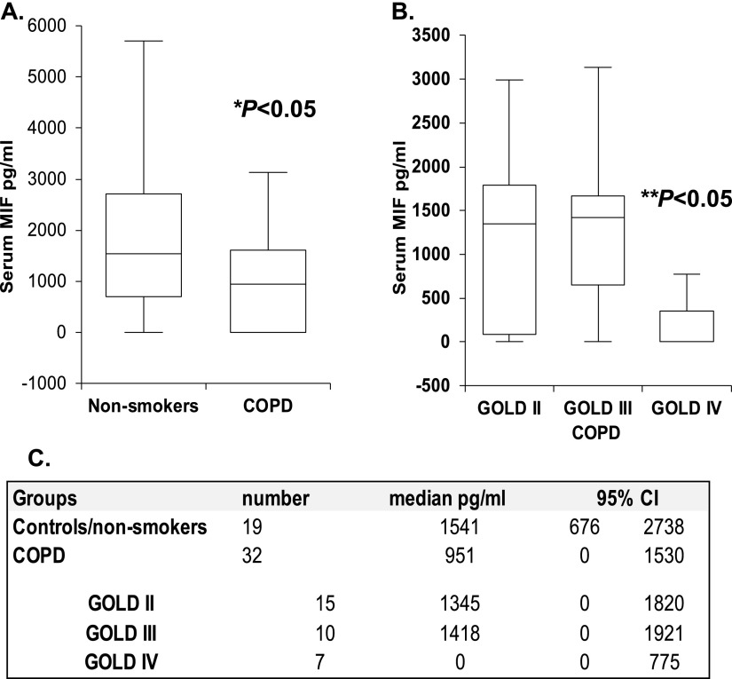
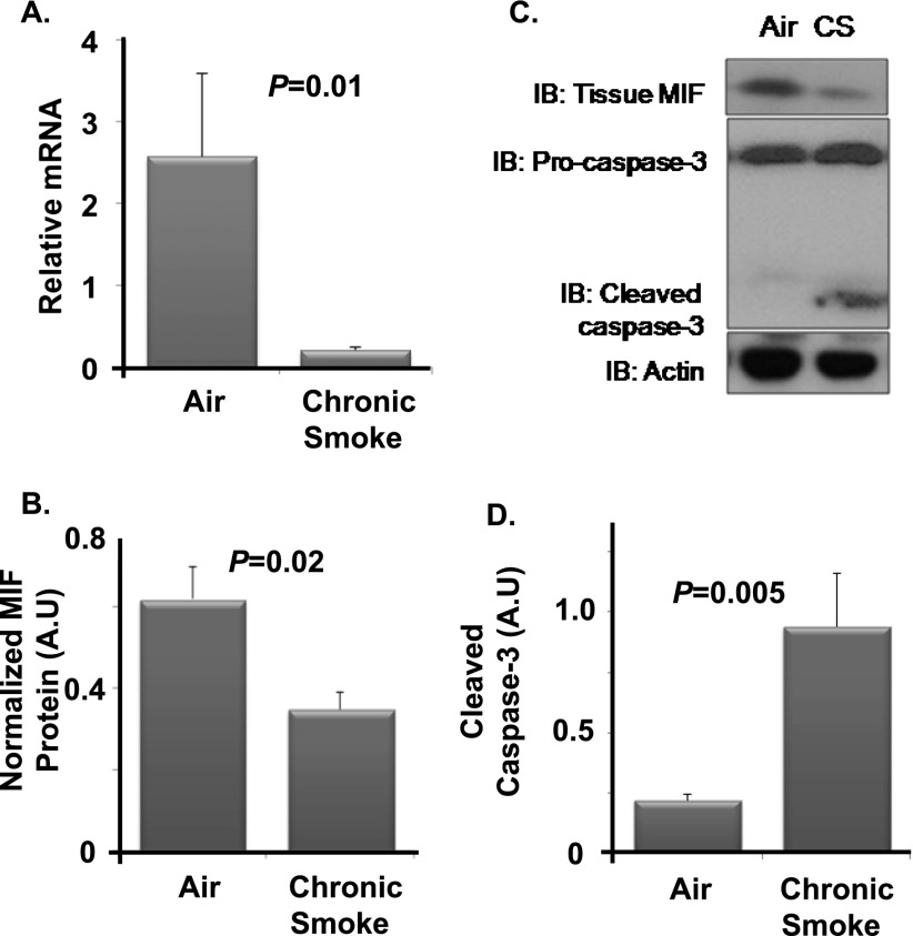
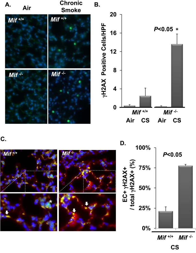
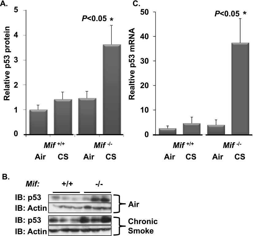
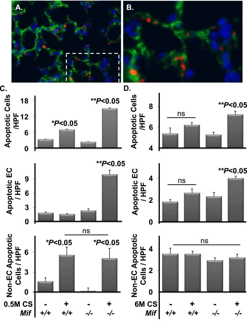
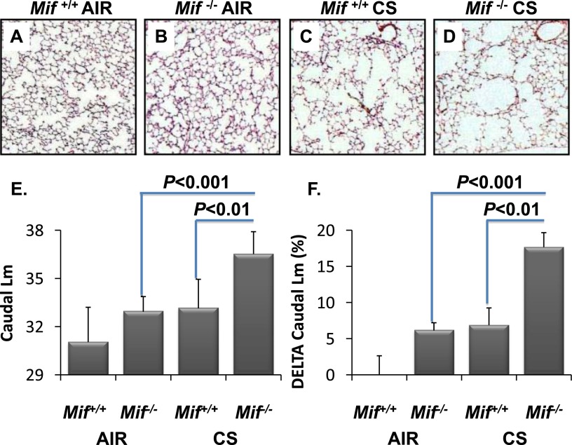
Cigarette smoke (CS) is the most common cause of chronic obstructive pulmonary diseases (COPD), including emphysema. CS exposure impacts all cell types within the airways and lung parenchyma, causing alveolar tissue destruction through four mechanisms: (1) oxidative stress; (2) inflammation; (3) protease-induced degradation of the extracellular matrix; and (4) enhanced alveolar epithelial and endothelial cell (EC) apoptosis. Studies in human pulmonary ECs demonstrate that macrophage migration inhibitory factor (MIF) antagonizes CS-induced apoptosis. Here, we used human microvascular ECs, an animal model of emphysema (mice challenged with chronic CS), and patient serum samples to address both the capacity of CS to alter MIF expression and the effects of MIF on disease severity. We demonstrate significantly reduced serum MIF levels in patients with COPD. In the murine model, chronic CS exposure resulted in decreased MIF mRNA and protein expression in the intact lung. MIF deficiency (Mif(-/-)) potentiated the toxicity of CS exposure in vivo via increased apoptosis of ECs, resulting in enhanced CS-induced tissue remodeling. This was linked to MIF's capacity to protect against double-stranded DNA damage and suppress p53 expression. Taken together, MIF appears to antagonize CS-induced toxicity in the lung and resultant emphysematous tissue remodeling by suppressing EC DNA damage and controlling p53-mediated apoptosis, highlighting a critical role of MIF in EC homeostasis within the lung.

Keywords: apoptosis; cigarette; emphysema; endothelial; macrophage migration inhibitory factor.


Figures

Figure 1.
Figure 1.
Serum macrophage migration inhibitory factor (MIF) concentrations are reduced in patients with severe chronic obstructive pulmonary diseases (COPD). Serum MIF levels were significantly reduced in subjects with COPD when compared with nonsmokers (A). In patients with COPD, serum MIF was significantly reduced in severe, GOLD (Global Initiative for Chronic Obstructive Lung Disease) IV disease (B). Values are expressed as median and 95% confidence interval (CI) (C). *P < 0.05, nonsmokers versus COPD; **P < 0.05, GOLD IV versus GOLD II and III.
Figure 2.
Figure 2.
Chronic smoke alters MIF expression in the intact lung. Wild-type C57BL/6 mice were exposed to cigarette smoke (CS) or filtered air. Gene expression and Western blotting were used to study alterations in MIF and cleaved caspase-3. Lung MIF mRNA was significantly reduced after exposure to CS (A). Tissue MIF was significantly decreased with chronic CS relative to air controls (B). The resulting protein intensities were normalized to β-actin (C). Chronic CS was associated with enhanced cleavage of caspase-3 relative to air controls (D). n = 5 per arm. Values are expressed as means ± SEM.
Figure 3.
Figure 3.
MIF antagonizes CS-induced endothelial cell (EC) double-stranded DNA breaks (DSBs) in vivo. Lungs from Mif+/+ and Mif−/− animals exposed to filtered air or CS for 0.5 months were harvested and sectioned for immunohistochemistry (A and C). MIF-deficient mice exposed to CS demonstrated a significant increase in γH2AX-positive parenchymal cells relative to air-exposed Mif−/− and CS-challenged Mif+/+ mice (A and B). Mif−/− mice had significantly higher γH2AX-positive (green) ECs as detected by colocalization of isolectin staining (red) (C and D). Area of magnification denoted with dashed lines (C). Arrow identifies double positive cells, * identifies H2AX positive/isolectin negative cells. n = 5 per arm. Values are expressed as means ± SEM. A.U., arbitrary unit; IB, immunoblot.
Figure 4.
Figure 4.
CS-induced p53 expression is increased in the absence of MIF. Lungs from Mif+/+ and Mif−/− animals exposed to filtered air or CS for 0.5 months were harvested and homogenized for Western blotting (A and B) and gene expression (C). Relative p53 protein expression was increased in Mif−/− mice exposed to CS (A). Representative Western blot (B). n = 5–6 per arm. There was a significant increase in p53 mRNA in Mif−/− animals in response to CS as assessed by comparative quantitative PCR (C). *P < 0.05 Mif−/ CS versus air and Mif+/+ CS.
Figure 5.
Figure 5.
Caspase-3 expression is increased in the absence of MIF with subacute and chronic CS exposure. Lungs from Mif+/+ and Mif−/− animals exposed to filtered air or CS for 0.5 or 6 months were harvested and sectioned for immunohistochemistry. Representative fluorescent microscopy images are shown of cleaved caspase-3 (red) and thrombomodulin (green) in the lung (A and B). Area of magnification denoted with dashed lines (A). The frequency of cleaved caspase-3–positive parenchymal cells was significantly increased in Mif−/− versus Mif+/+ mice exposed to 0.5 months of CS (P < 0.05) ([C] upper panel), and with 6 months of CS (P < 0.05) ([D] upper panel). The majority of caspase-3–positive cells in Mif−/−exposed to 0.5 or 6 months of CS were ECs (both with **P < 0.05) (middle panels [C and D]). Non-ECs were enhanced with 0.5 months with exposure (*P < 0.05), and this did not differ with genoptype (lower panels [C and D]). No differences were observed under basal conditions independent of genotype (n = 5–8 per arm). Nuclei were stained with 4′,6-diamidino-2-phenylindole (blue). Values are expressed as means ± SEM. ns, not significant. *P < 0.05 air versus CS. **P < 0.05 Mif+/+ versus Mif−/−.
Figure 6.
Figure 6.
MIF deficiency leads to increased sensitivity to emphysematous remodeling in vivo. Lungs from Mif+/+ and Mif−/− animals exposed to filtered air or CS for 6 months were sectioned for morphometry. Differences in alveolar remodeling in Mif+/+ versus Mif−/− mice are visually apparent in CS-exposed caudal lung regions (C and D, respectively) compared with air-exposed mice (A and B, respectively). Mif−/− mice had significantly higher mean chord length (Lm) in the caudal lung regions than Mif+/+ when exposed to CS (E), and the change in Lm from baseline was significant in Mif−/− mice (F). Values are expressed as means ± SEM.

References

    1. Gordon C, Gudi K, Krause A, Sackrowitz R, Harvey BG, Strulovici-Barel Y, Mezey JG, Crystal RG. Circulating endothelial microparticles as a measure of early lung destruction in cigarette smokers. Am J Respir Crit Care Med. 2011;184:224–232. - PMC - PubMed
    1. Henson PM, Cosgrove GP, Vandivier RW. State of the art: apoptosis and cell homeostasis in chronic obstructive pulmonary disease. Proc Am Thorac Soc. 2006;3:512–516. - PMC - PubMed
    1. Kanazawa H, Yoshikawa J. Elevated oxidative stress and reciprocal reduction of vascular endothelial growth factor levels with severity of COPD. Chest. 2005;128:3191–3197. - PubMed
    1. Calabrese F, Giacometti C, Beghe B, Rea F, Loy M, Zuin R, Marulli G, Baraldo S, Saetta M, Valente M. Marked alveolar apoptosis/proliferation imbalance in end-stage emphysema. Respir Res. 2005;6:14. - PMC - PubMed
    1. Giordano RJ, Lahdenranta J, Zhen L, Chukwueke U, Petrache I, Langley RR, Fidler IJ, Pasqualini R, Tuder RM, Arap W. Targeted induction of lung endothelial cell apoptosis causes emphysema-like changes in the mouse. J Biol Chem. 2008;283:29447–29460. - PMC - PubMed

Publication types

MeSH terms