. 2014 Apr 18;9(4):967-75.
doi: 10.1021/cb500026x. Epub 2014 Feb 11.

De novo design of self-assembling foldamers that inhibit heparin-protein interactions

Affiliations

De novo design of self-assembling foldamers that inhibit heparin-protein interactions

Geronda L Montalvo et al. ACS Chem Biol. .

Abstract

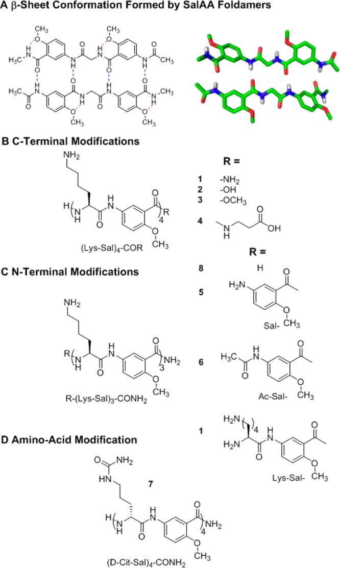
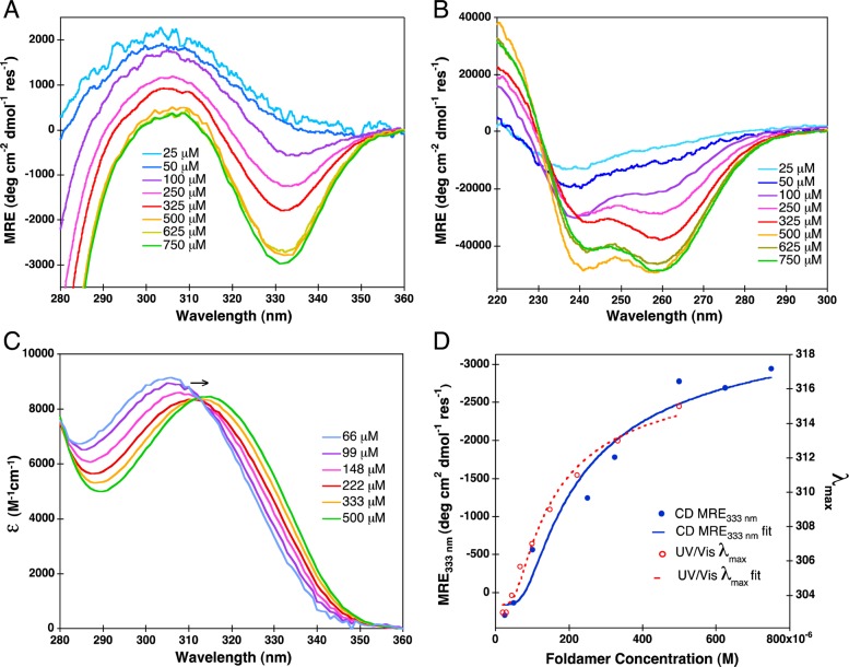
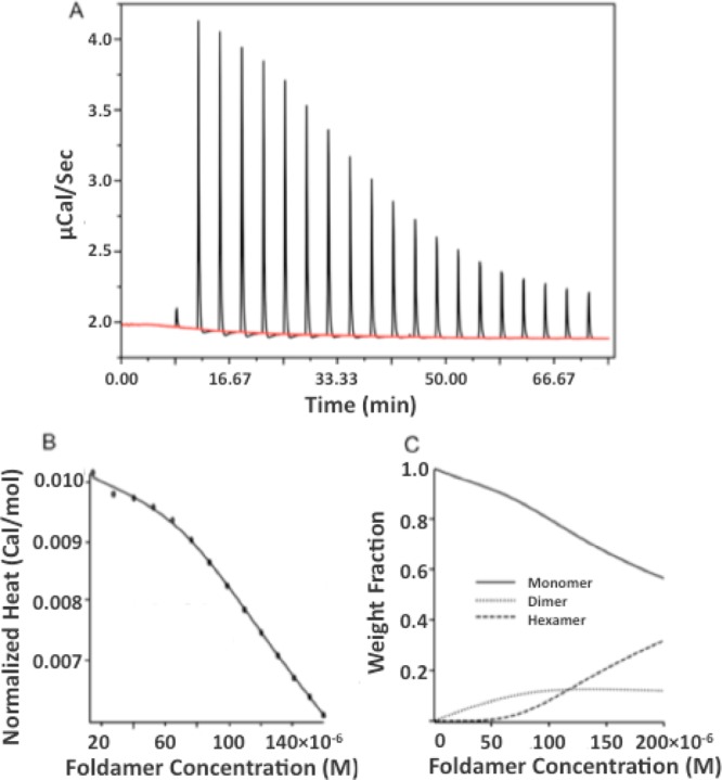
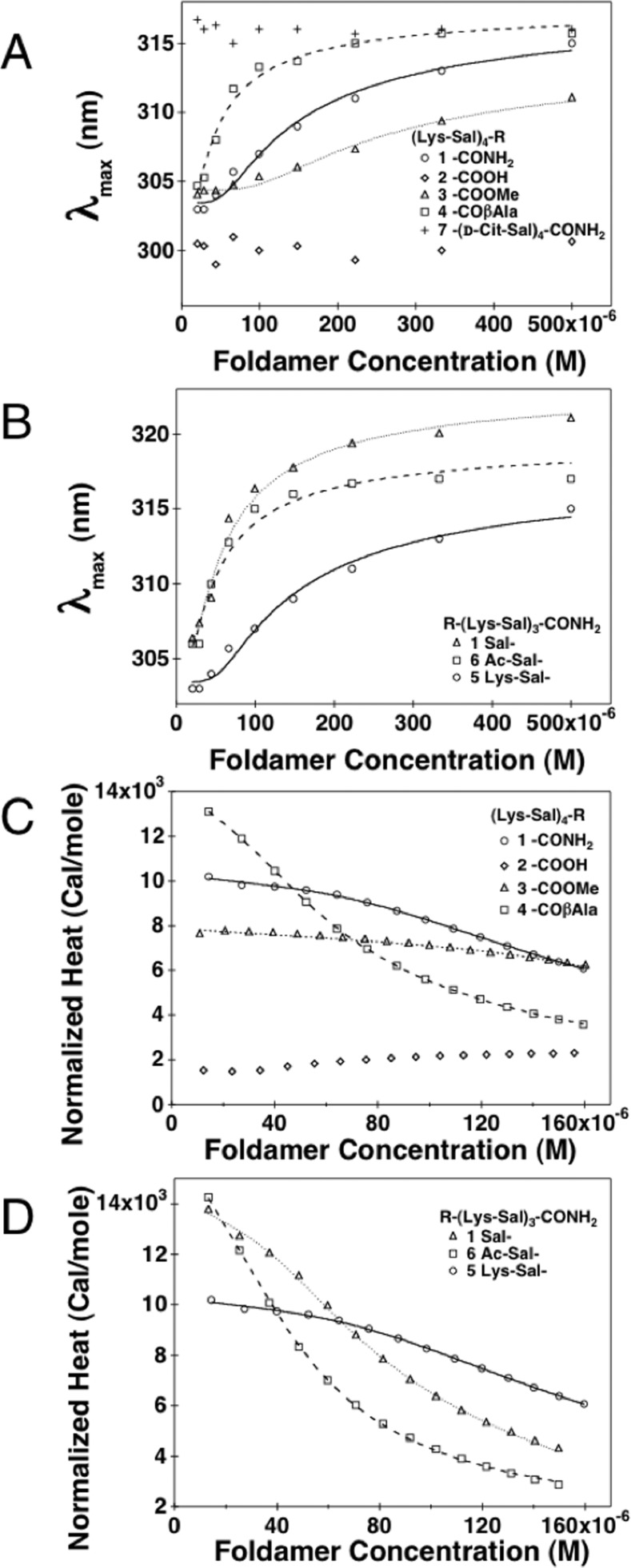
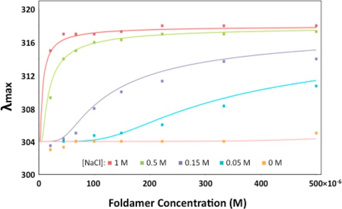
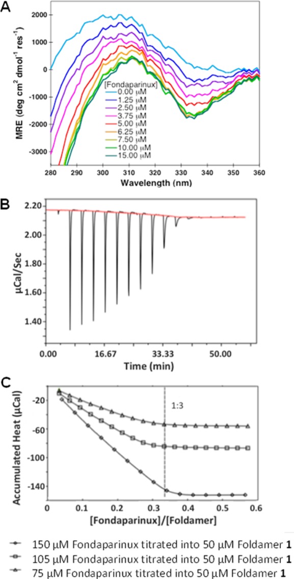
A series of self-associating foldamers have been designed as heparin reversal agents, as antidotes to prevent bleeding due to this potent antithrombotic agent. The foldamers have a repeating sequence of Lys-Sal, in which Sal is 5-amino-2-methoxy-benzoic acid. These foldamers are designed to self-associate along one face of an extended chain in a β-sheet-like interaction. The methoxy groups were included to form intramolecular hydrogen bonds that preclude the formation of very large amyloid-like aggregates, while the positively charged Lys side chains were introduced to interact electrostatically with the highly anionic heparin polymer. The prototype compound (Lys-Sal)4 carboxamide weakly associates in aqueous solution at physiological salt concentration in a monomer-dimer-hexamer equilibrium. The association is greatly enhanced at either high ionic strength or in the presence of a heparin derivative, which is bound tightly. Variants of this foldamer are active in an antithrombin III-factor Xa assay, showing their potential as heparin reversal agents.


Figures

Figure 1
Figure 1
Chemical structures of SalAA foldamers explored in this study. (A) Schematic (left) and crystal structure (right) of a dimeric foldamer based on the salicylamide backbone, Ac-Sal-Gly-Sal-NMe amide, in which Ac is acetyl, Sal is the indicated amino-salicylic acid methyl ether unit, Gly is glycine, and NMe amide is an N-methyl amide. (B) Chemical structures of salicylamide-α-amino acid peptides (SalAA foldamers) for C-terminal and N-terminal (C) modified series. (D) Chemical structure of compound 7, (d-Cit-Sal)4-CONH2; in this variant of (Lys-Sal)4-CONH2, compound 1, cationic lysine is replaced with a neutral citrulline amino acid.
Figure 2
Figure 2
Concentration-dependent CD spectra of compound 1 in the near (A) and far (B) UV regions. Extinction coefficient as a function of wavelength (C), determined from near UV–vis absorption spectra. CD mean residue ellipticity (MRE) at 333 nm and UV–vis absorption shifts in λmax are overlaid (D) with their respective fits; Khex equals 10–18.4 (or 4.0 × 10⁻¹⁹) M5 for CD MRE333 and 10–19.3 (or 5 × 10–20) M5 for UV–vis λmax.
Figure 3
Figure 3
Concentration-dependent ITC of 1. A stock solution of 1.0 mM 1 in TBS was serially diluted into the same buffer (A). The heat associated with each injection was determined by integrating the peaks. The heat generated for a given final concentration is then summed and expressed in cal/mol vs final concentration (B). The smooth line shows the best fit of a monomer-dimer-hexamer equilibrium to the data (see Methods). The weight fraction distribution for hexamer, dimer, and monomer species are plotted vs concentration (C).
Figure 4
Figure 4
UV–vis absorption λmax and normalized ITC heats of dilution as a function of SalAA foldamer concentration. (A) UV–vis absorption λmax as a function of SalAA foldamer concentration for C-terminal modifications. (B) UV–vis absorption λmax as a function of SalAA foldamer concentration for N-terminal modifications. Normalized ITC heats of dilution (pK(hex-mon)) values are summarized in Table 2 as a function of SalAA foldamer concentration for C-terminal (C) and N-terminal (D) modified series, and their respective fits are illustrated as lines.
Figure 5
Figure 5
Concentration-dependent shifts in λmax for 1 at varying [NaCl] in 0.05 M Tris, pH 7.4. The smooth curves were generated by global fitting of the data to a monomer-hexamer equilibrium. The value of λmax for the monomer (304 nm) and hexamer (318 nm) were globally fit, while the value of pK(hex-mon) was allowed to vary for each curve. The fit pK(hex-mon) values were <15 at 0 M NaCl, 17.4 at 0.05 M, 19.7 at 0.15 M, 24.1 at 0.5 M, and 26.5 at 1.0 M.
Figure 6
Figure 6
Near UV CD and ITC characterizations of SalAA compound 1 binding to fondaparinux. (A) Near UV CD spectra of the fondaparinux titration of compound 1, (Lys-Sal)4-CONH2, where increasing concentrations of fondaparinux were titrated into 25 μM compound 1. (B) Concentration-dependent ITC of fondaparinux titrated into compound 1, (Lys-Sal)4-CONH2. Shown are the raw data of 150 μM fondaparinux (stock solution) titrated into 50 μM compound 1. (C) Accumulated integrated heat changes as a function of fondaparinux:foldamer molar ratio for three experiments at the listed concentrations.

References

    1. Gellman S. H. (1998) Foldamers: a manifesto. Acc. Chem. Res. 31, 173–180.
    1. Goodman C. M.; Choi S.; Shandler S.; DeGrado W. F. (2007) Foldamers as versatile frameworks for the design and evolution of function. Nat. Chem. Biol. 3, 252–262. - PMC - PubMed
    1. Guichard G.; Huc I. (2011) Synthetic foldamers. Chem. Commun. 47, 5933–5941. - PubMed
    1. Hill D. J.; Mio M. J.; Prince R. B.; Hughes T. S.; Moore J. S. (2001) A field guide to foldamers. Chem. Rev. 101, 3893–4012. - PubMed
    1. Choi S.; Clements D. J.; Pophristic V.; Ivanov I.; Vemparala S.; Bennett J. S.; Klein M. L.; Winkler J. D.; DeGrado W. F. (2005) The design and evaluation of heparin-binding foldamers. Angew. Chem., Int. Ed. 44, 6685–6689. - PubMed

Publication types