

Acute murine antigen-induced arthritis is not affected by disruption of osteoblastic glucocorticoid signalling
- PMID: 24491163
- PMCID: PMC3922092
- DOI: 10.1186/1471-2474-15-31
Acute murine antigen-induced arthritis is not affected by disruption of osteoblastic glucocorticoid signalling
Abstract
Background: The role of endogenous glucocorticoids (GC) in the initiation and maintenance of rheumatoid arthritis (RA) remains unclear. We demonstrated previously that disruption of GC signalling in osteoblasts results in a profound attenuation of K/BxN serum-induced arthritis, a mouse model of RA. To determine whether or not the modulation of the inflammatory response by osteoblasts involves T cells, we studied the effects of disrupted osteoblastic GC-signalling in the T cell-dependent model of antigen-induced arthritis (AIA).
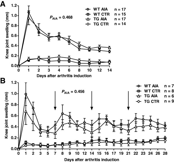
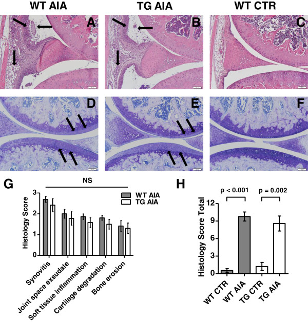
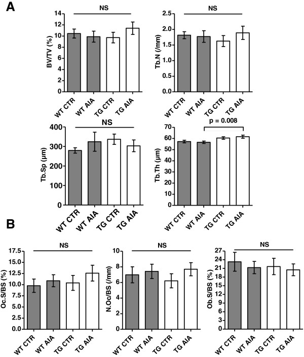
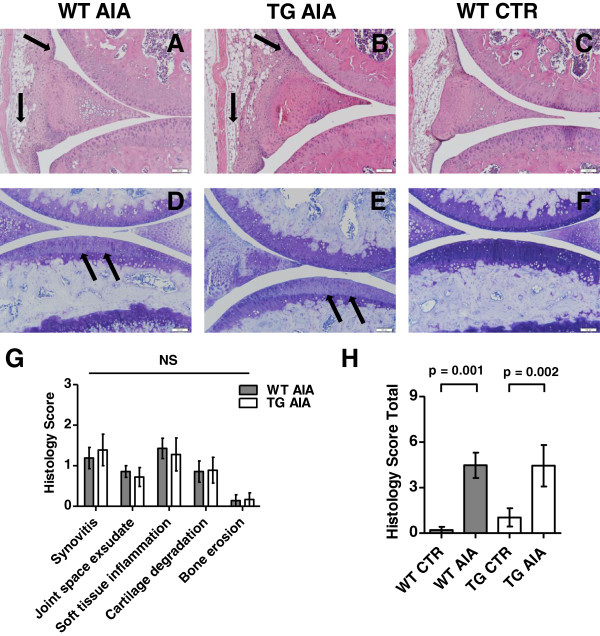
Methods: Acute arthritis was induced in pre-immunised 11-week-old male 11β-hydroxysteroid dehydrogenase type 2 transgenic (tg) mice and their wild-type (WT) littermates by intra-articular injection of methylated bovine serum albumine (mBSA) into one knee joint. Knee diameter was measured every 1-2 days until euthanasia on day 14 post injection. In a separate experiment, arthritis was maintained for 28 days by weekly reinjections of mBSA. Tissues were analysed by histology, histomorphometry and microfocal-computed tomography. Serum cytokines levels were determined by multiplex suspension array.
Results: In both short and long term experiments, arthritis developed in tg and WT mice with no significant difference between both groups. Histological indices of inflammation, cartilage damage and bone erosion were similar in tg and WT mice. Bone volume and turnover at the contralateral tibia and systemic cytokine levels were not different.
Conclusions: Acute murine AIA is not affected by a disruption in osteoblastic GC signalling. These data indicate that osteoblasts do not modulate the T cell-mediated inflammatory response via a GC-dependent pathway.
Figures
Similar articles
-
Transgenic disruption of endogenous glucocorticoid signaling in osteoblasts does not alter long-term K/BxN serum transfer-induced arthritis.Arthritis Res Ther. 2023 Aug 4;25(1):140. doi: 10.1186/s13075-023-03112-9. Arthritis Res Ther. 2023. PMID: 37542341 Free PMC article.
-
Arthritis and the role of endogenous glucocorticoids.Bone Res. 2020 Sep 8;8:33. doi: 10.1038/s41413-020-00112-2. eCollection 2020. Bone Res. 2020. PMID: 32963891 Free PMC article. Review.
-
Transgenic disruption of glucocorticoid signaling in mature osteoblasts and osteocytes attenuates K/BxN mouse serum-induced arthritis in vivo.Arthritis Rheum. 2009 Jul;60(7):1998-2007. doi: 10.1002/art.24619. Arthritis Rheum. 2009. PMID: 19565501
-
Transgenic Disruption of Glucocorticoid Signaling in Osteoblasts Attenuates Joint Inflammation in Collagen Antibody-Induced Arthritis.Am J Pathol. 2016 May;186(5):1293-301. doi: 10.1016/j.ajpath.2015.12.025. Epub 2016 Mar 14. Am J Pathol. 2016. PMID: 26988651
-
Endogenous glucocorticoid signaling in chondrocytes attenuates joint inflammation and damage.FASEB J. 2018 Jan;32(1):478-487. doi: 10.1096/fj.201700659R. Epub 2017 Sep 19. FASEB J. 2018. PMID: 28928247
Cited by
-
[Osteoimmunology-IMMUNOBONE : Regulation of bone by inflammation].Z Rheumatol. 2018 May;77(Suppl 1):12-15. doi: 10.1007/s00393-018-0455-0. Z Rheumatol. 2018. PMID: 29691689 Review. German. No abstract available.
-
Secondary osteoporosis in collagen-induced arthritis rats.J Bone Miner Metab. 2016 Sep;34(5):500-16. doi: 10.1007/s00774-015-0700-4. Epub 2015 Jul 26. J Bone Miner Metab. 2016. PMID: 26210858
-
Transgenic disruption of endogenous glucocorticoid signaling in osteoblasts does not alter long-term K/BxN serum transfer-induced arthritis.Arthritis Res Ther. 2023 Aug 4;25(1):140. doi: 10.1186/s13075-023-03112-9. Arthritis Res Ther. 2023. PMID: 37542341 Free PMC article.
-
Arthritis and the role of endogenous glucocorticoids.Bone Res. 2020 Sep 8;8:33. doi: 10.1038/s41413-020-00112-2. eCollection 2020. Bone Res. 2020. PMID: 32963891 Free PMC article. Review.
-
Preventive effects of Polygonum multiflorum on glucocorticoid-induced osteoporosis in rats.Exp Ther Med. 2017 Sep;14(3):2445-2460. doi: 10.3892/etm.2017.4802. Epub 2017 Jul 18. Exp Ther Med. 2017. PMID: 28962180 Free PMC article.
References
-
- Buttgereit F, Zhou H, Seibel MJ. Arthritis and endogenous glucocorticoids: the emerging role of the 11beta-HSD enzymes. Ann Rheum Dis. 2008;67(9):1201–1203. - PubMed
-
- Saldanha C, Tougas G, Grace E. Evidence for anti-inflammatory effect of normal circulating plasma cortisol. Clin Exp Rheumatol. 1986;4(4):365–366. - PubMed
-
- Coutinho AE, Gray M, Brownstein DG, Salter DM, Sawatzky DA, Clay S, Gilmour JS, Seckl JR, Savill JS, Chapman KE. 11beta-Hydroxysteroid dehydrogenase type 1, but not type 2, deficiency worsens acute inflammation and experimental arthritis in mice. Endocrinology. 2012;153(1):234–240. doi: 10.1210/en.2011-1398. - DOI - PMC - PubMed
Publication types
MeSH terms
Substances
LinkOut - more resources
Full Text Sources
Other Literature Sources
Medical
Miscellaneous
