. 2014 Apr;24(2):139-48.
doi: 10.1089/nat.2013.0465. Epub 2014 Feb 3.

Quantum mechanical studies of DNA and LNA

Affiliations

Quantum mechanical studies of DNA and LNA

Troels Koch et al. Nucleic Acid Ther. 2014 Apr.

Abstract

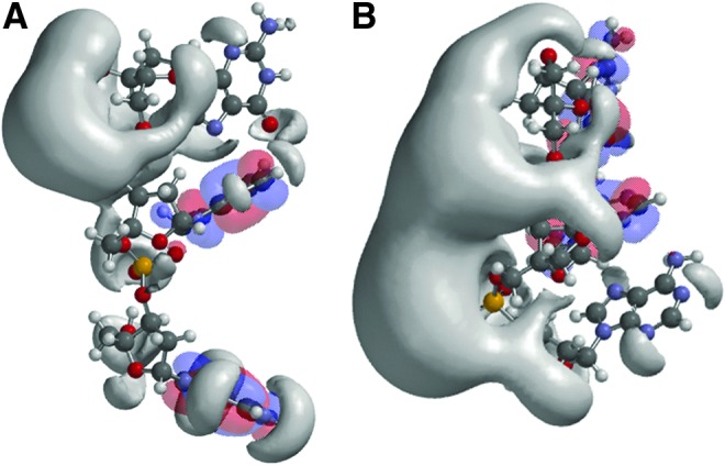
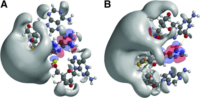
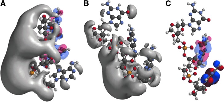
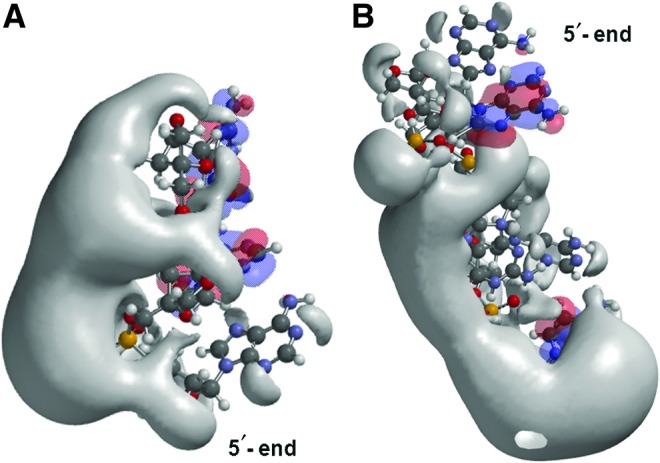
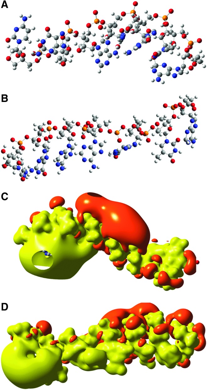
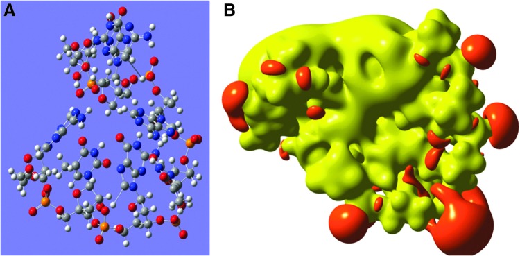
Quantum mechanical (QM) methodology has been employed to study the structure activity relations of DNA and locked nucleic acid (LNA). The QM calculations provide the basis for construction of molecular structure and electrostatic surface potentials from molecular orbitals. The topologies of the electrostatic potentials were compared among model oligonucleotides, and it was observed that small structural modifications induce global changes in the molecular structure and surface potentials. Since ligand structure and electrostatic potential complementarity with a receptor is a determinant for the bonding pattern between molecules, minor chemical modifications may have profound changes in the interaction profiles of oligonucleotides, possibly leading to changes in pharmacological properties. The QM modeling data can be used to understand earlier observations of antisense oligonucleotide properties, that is, the observation that small structural changes in oligonucleotide composition may lead to dramatic shifts in phenotypes. These observations should be taken into account in future oligonucleotide drug discovery, and by focusing more on non RNA target interactions it should be possible to utilize the exhibited property diversity of oligonucleotides to produce improved antisense drugs.


Figures

<b>Fig. 1.</b>
Fig. 1.
Structures and frontier orbitals (red/blue) for (A) DNA and (B) LNA with the sequence 5′-AAG-3′ (5′ end at bottom of structure). The electrostatic potential (negative) is shown in gray and represented by an Iso-potential value of −83 kJ/mol.
<b>Fig. 2.</b>
Fig. 2.
Structures and frontier orbitals (red/blue) for LNA trinucleotides with sequence 5′-AAG-3′, chemically modified by phosphorothioate internucleoside linkages (PS). In (A), the configuration of PS is RS and in (B) the configuration is SR. The 5′ end is at the top in A and at the bottom in B. The electrostatic potential (negative) is shown in gray and represented by an Iso-potential value of −83 kJ/mol.
<b>Fig. 3.</b>
Fig. 3.
Structures and frontier orbitals (red/blue) for LNA tri-nucleotides with sequence 5′-AAG-3′ (A) and 5′-AGG-3′ (B). The 5′ end is at the bottom of the figure. In (C) the HOMO-LUMO orbitals are explicitly shown for LNA 5′-AGG-3′. The electrostatic potential (negative) is shown in gray and represented by an Iso-potential value of −83 kJ/mol.
<b>Fig. 4.</b>
Fig. 4.
Structures and frontier orbitals (red/blue) for (A) LNA tri-nucleotide with sequence 5′-AAG-3′, and (B) LNA penta-nucleotide with sequence 5′-AAGAC-3′. The 5′ end is at the bottom in (A) and is at the top in (B). The electrostatic potential (negative) is shown in gray and represented by an Iso-potential value of −83 kJ/mol.
<b>Fig. 5.</b>
Fig. 5.
Three-dimensional chemical structures of the LNA heptanucleotides (A) 5′-ATGTAGC-3′ and (B) 5′-ATGAAGC-3′. The 5′ end is placed to the right. The topology of the electrostatic potentials of the two heptanucleotides is shown in (C) 5′-ATGTAGC-3′ and (D) 5′-ATGAAGC-3′. The energy of the electrostatic iso-potential surfaces is set to 1.63 eV (157 kJ/mol). The negative potential is marked with red and the positive with yellow. The 5′ end is to the right.
<b>Fig. 6.</b>
Fig. 6.
Three-dimensional chemical structure of the LNA-DNA PO gap-mer 5′-ATGTAGC-3′ (LNA nts underlined) (A), and in (B) the electrostatic potential represented by an iso-surface value of iso=0.06H=1.62eV=156 kJ/mol. The positive electrostatic potential is shown in yellow and the negative in red.
<b>Fig. 7.</b>
Fig. 7.
Illustration of “extreme properties” of drug candidates. Most oligonucleotides tend to cluster around a central value for each variable (phenotype) in search. The dashed circles illustrate the atypical and extreme properties that are selected for in drug discovery: high pharmacological activity combined with low toxicity.

References

    1. ABDALI S., JALKANEN K.J., BOHR H., SUHAI S., and NIEMINEN R.M. (2002). The VA and VCD spectra of various isotopes of L-alanine in aqueous solution. Chem. Phys. 282,219–235
    1. ALTMANN K.-H., DEAN N.M., FABBRO D., FREIER S.M., GEIGER T., HÄNER R., HÜSKEN D., MARTIN P., MONIA B.P., MÜLLER M., et al. (1996). Second generation OD antisense oligonucleotides: from nuclease resistance to biological efficacy in animals. Chimia 50,168–176
    1. BELTINGER C., SARAGOVI H.U., SMITH R.M., LESAUTEUR L., SHAH N., DEDIONISIO L., CHRISTENSEN L., RAIBLE A., JARETT L., and GEWIRTZ A.M. (1995). Binding, uptake, and intracellular trafficking of phosphorothioate-modified oligodeoxynucleotides. J. Clin. Invest 95,1814–1823 - PMC - PubMed
    1. BOHR H.G. (2013). Perspectives in quantum nanobiology and biophysical chemistry. Current Phys. Chem. 3,4–8
    1. BURGOYNE N.J., and JACKSON R.M. (2006). Predicting protein interaction sites: binding hot-spots in protein-protein and protein-ligand interfaces. Bioinformatics 22,1335–1342 - PubMed