Review
. 2014 May;20(5):251-60.
doi: 10.1016/j.molmed.2014.01.004. Epub 2014 Jan 31.

Polycystin-1: a master regulator of intersecting cystic pathways

Affiliations
Review

Polycystin-1: a master regulator of intersecting cystic pathways

Sorin V Fedeles et al. Trends Mol Med. 2014 May.

Abstract

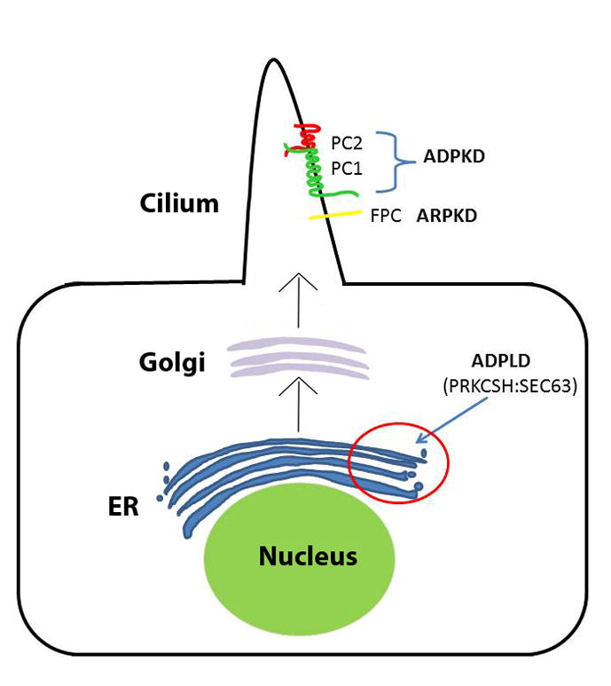
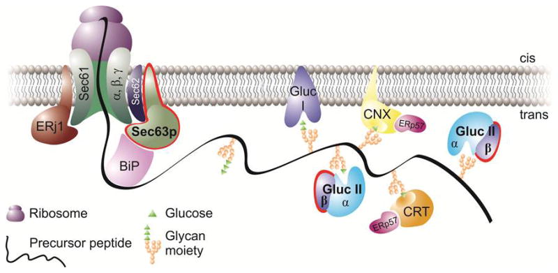
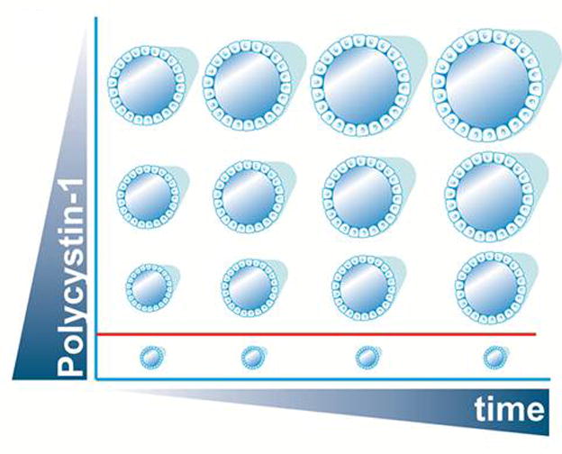
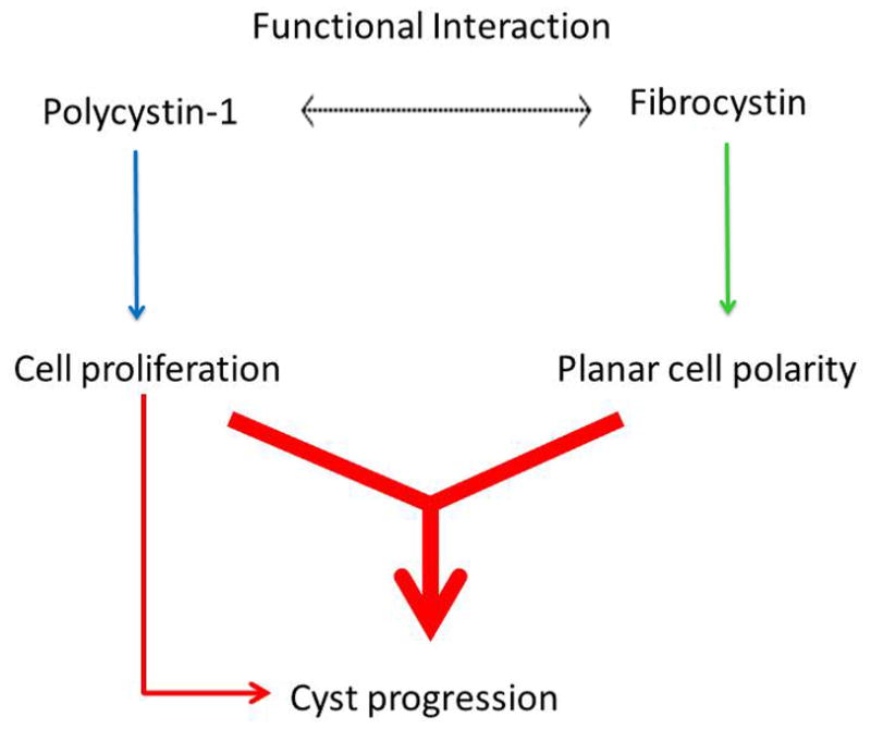
Autosomal dominant polycystic kidney disease (ADPKD) is the most common potentially lethal monogenic disorder, with more than 12 million cases worldwide. The two causative genes for ADPKD, PKD1 and PKD2, encode protein products polycystin-1 (PC1) and polycystin-2 (PC2 or TRPP2), respectively. Recent data have shed light on the role of PC1 in regulating the severity of the cystic phenotypes in ADPKD, autosomal recessive polycystic kidney disease (ARPKD), and isolated autosomal dominant polycystic liver disease (ADPLD). These studies showed that the rate for cyst growth was a regulated trait, a process that can be either sped up or slowed down by alterations in functional PC1. These findings redefine the previous understanding that cyst formation occurs as an 'on-off' process. Here, we review these and other related studies with an emphasis on their translational implications for polycystic diseases.

Keywords: chaperone therapy; cyst progression; polycystic kidney disease; polycystic liver disease; polycystin-1 dosage; protein biogenesis.


Figures

Figure 1
Figure 1
Schematic view of the relevant subcellular localization of the proteins involved in Autosomal Dominant Polycystic Kidney Disease (ADPKD), Autosomal Recessive Polycystic Kidney Disease (ARPKD), and Autosomal Dominant Polycystic Liver Disease (ADPLD). The two ADPLD proteins glucosidase IIβ (GIIβ) and Sec63p are localized in the ER and are involved in protein translocation (Sec63p) and folding (GIIβ). The proteins associated with ADPKD, polycystin-1 (PC-1, encoded by PKD1) and polycystin-2 (PC-2, encoded by PKD2), in addition to the ARPKD protein fibrocystin/polyductin, (FPC, encoded by PKHD1) are all ciliary transmembrane proteins which need to go through the co-translational translocation pathway (see Figure 2) in the endoplasmic reticulum (ER), which Sec63p and GIIβ are integral members of, in order to be properly folded and trafficked to Golgi and onto their final physiological destination, the primary cilia.
Figure 2
Figure 2
The co-translational translocation pathway highlighting (red contour) the two ADPLD proteins, Sec63p and GIIβ. At this stage, the precursor protein (e.g. PC1) is threaded through the translocon, with its central subunit, the Sec61 complex, and the auxiliary subunits that are present in the membrane (Sec63 and ERdj1/Erj1). Via their DnaJ domains, Sec63p and Erdj1 provide a link to the ATPase cycle of the luminal chaperone BiP. Downstream of Sec63p, GIIβ is involved (as the regulatory subunit of the glucose trimming enzyme, glucosidase II) in glucose trimming, which takes place as part of the calnexin-calreticulin cycle. Cystic proteins such as PC1 have to go through this pathway to achieve their final, functional cellular location (the primary cilia). Defects along this pathway may affect both the translocation (due to Sec63p inactivation) or glucose trimming/folding (via GIIβ) of cystic proteins as described in [20].
Figure 3
Figure 3
Schematic view showing that PC1 levels can dictate the timing and severity of the phenotype in a time (x-axis) and dose (y-axis) dependent manner. For a certain PC1 threshold (depicted by the red line) no cyst formation occurs. When the dose of PC1 goes below this threshold, cyst initiation/progression ensues with lower levels leading to faster cyst growth. This is evidenced by the ADPLD mouse models where the lower dosage of PC1 on the Sec63 deficient background leads to an earlier manifestation of the phenotype as compared to the Prkcsh null background.
Figure 4
Figure 4
Diagram of the genetic interaction between Pkd1 and the ARPKD locus. Fibrocystin plays a role in cyst progression through its involvement in the planar cell polarity pathway (green arrow). Polycystin-1 (PC1), through its link to cell proliferation (blue arrow), can further contribute to the severity of the phenotype, as seen in the double Pkd1/Pkhd1 mutants. Since defects in planar cell polarity in the absence of Pkhd1 do not lead to polycystic kidney disease, the contribution of planar cell polarity to cyst progression occurs in the context of increased proliferation (red arrow). Is it not clear what the functional interaction between PC1 and Fibrocystin (dotted arrow) is, for example whether there is a direct biochemical connection between the two proteins impacting their respective function/ levels.

References

    1. Guay-Woodford LM. Renal cystic diseases: diverse phenotypes converge on the cilium/centrosome complex. Pediatr Nephrol. 2006;21:1369–1376. - PubMed
    1. Hughes J, et al. The polycystic kidney disease 1 (PKD1) gene encodes a novel protein with multiple cell recognition domains. Nat Genet. 1995;10:151–160. - PubMed
    1. Mochizuki T, et al. PKD2, a gene for polycystic kidney disease that encodes an integral membrane protein. Science. 1996;272:1339–1342. - PubMed
    1. Nauli SM, et al. Polycystins 1 and 2 mediate mechanosensation in the primary cilium of kidney cells. Nat Genet. 2003;33:129–137. - PubMed
    1. Qian F, et al. The molecular basis of focal cyst formation in human autosomal dominant polycystic kidney disease type I. Cell. 1996;87:979–987. - PubMed

Publication types

Substances