Olfactory maps, circuits and computations
- PMID: 24492088
- PMCID: PMC3913910
- DOI: 10.1016/j.conb.2013.09.010
Olfactory maps, circuits and computations
Abstract
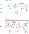
Sensory information in the visual, auditory and somatosensory systems is organized topographically, with key sensory features ordered in space across neural sheets. Despite the existence of a spatially stereotyped map of odor identity within the olfactory bulb, it is unclear whether the higher olfactory cortex uses topography to organize information about smells. Here, we review recent work on the anatomy, microcircuitry and neuromodulation of two higher-order olfactory areas: the piriform cortex and the olfactory tubercle. The piriform is an archicortical region with an extensive local associational network that constructs representations of odor identity. The olfactory tubercle is an extension of the ventral striatum that may use reward-based learning rules to encode odor valence. We argue that in contrast to brain circuits for other sensory modalities, both the piriform and the olfactory tubercle largely discard any topography present in the bulb and instead use distributive afferent connectivity, local learning rules and input from neuromodulatory centers to build behaviorally relevant representations of olfactory stimuli.
Copyright © 2013 Elsevier Ltd. All rights reserved.
Figures


Similar articles
-
Distinct representations of olfactory information in different cortical centres.Nature. 2011 Apr 14;472(7342):213-6. doi: 10.1038/nature09868. Epub 2011 Mar 30. Nature. 2011. PMID: 21451525 Free PMC article.
-
Olfactory perception as a compass for olfactory neural maps.Trends Cogn Sci. 2011 Nov;15(11):537-45. doi: 10.1016/j.tics.2011.09.007. Epub 2011 Oct 14. Trends Cogn Sci. 2011. PMID: 22001868 Review.
-
Task-Demand-Dependent Neural Representation of Odor Information in the Olfactory Bulb and Posterior Piriform Cortex.J Neurosci. 2019 Dec 11;39(50):10002-10018. doi: 10.1523/JNEUROSCI.1234-19.2019. Epub 2019 Oct 31. J Neurosci. 2019. PMID: 31672791 Free PMC article.
-
Coding and transformations in the olfactory system.Annu Rev Neurosci. 2014;37:363-85. doi: 10.1146/annurev-neuro-071013-013941. Epub 2014 Jun 2. Annu Rev Neurosci. 2014. PMID: 24905594 Review.
-
The Olfactory Mosaic: Bringing an Olfactory Network Together for Odor Perception.Perception. 2017 Mar-Apr;46(3-4):320-332. doi: 10.1177/0301006616663216. Epub 2016 Sep 29. Perception. 2017. PMID: 27687814 Free PMC article. Review.
Cited by
-
The neurobiology of parenting and infant-evoked aggression.Physiol Rev. 2025 Jan 1;105(1):315-381. doi: 10.1152/physrev.00036.2023. Epub 2024 Aug 15. Physiol Rev. 2025. PMID: 39146250 Free PMC article. Review.
-
The regulatory landscape of retinoblastoma: a pathway analysis perspective.R Soc Open Sci. 2022 May 18;9(5):220031. doi: 10.1098/rsos.220031. eCollection 2022 May. R Soc Open Sci. 2022. PMID: 35620002 Free PMC article.
-
Organization of prefrontal network activity by respiration-related oscillations.Sci Rep. 2017 Mar 28;7:45508. doi: 10.1038/srep45508. Sci Rep. 2017. PMID: 28349959 Free PMC article.
-
Olfactory Optogenetics: Light Illuminates the Chemical Sensing Mechanisms of Biological Olfactory Systems.Biosensors (Basel). 2021 Aug 31;11(9):309. doi: 10.3390/bios11090309. Biosensors (Basel). 2021. PMID: 34562900 Free PMC article. Review.
-
Oxytocin Mediates Entrainment of Sensory Stimuli to Social Cues of Opposing Valence.Neuron. 2015 Jul 1;87(1):152-63. doi: 10.1016/j.neuron.2015.06.022. Neuron. 2015. PMID: 26139372 Free PMC article.
References
-
- Murthy VN. Olfactory maps in the brain. Annu Rev Neurosci. 2011;34:233–258. - PubMed
-
- Petersen CC. The functional organization of the barrel cortex. Neuron. 2007;56:339–355. - PubMed
-
- Luo L, Flanagan JG. Development of continuous and discrete neural maps. Neuron. 2007;56:284–300. - PubMed
-
- Layton OW, Mingolla E, Yazdanbakhsh A. Dynamic coding of border-ownership in visual cortex. J Vis. 2012;12:8. - PubMed
-
- Gonzalez F, Perez R. Neural mechanisms underlying stereoscopic vision. Prog Neurobiol. 1998;55:191–224. - PubMed
Publication types
MeSH terms
Grants and funding
LinkOut - more resources
Full Text Sources
Other Literature Sources

