Editorial
. 2014 Feb;15(2):175-6.
doi: 10.1097/PCC.0000000000000037.

Searching the right way to treat neonatal abstinence syndrome*

Affiliations
Editorial

Searching the right way to treat neonatal abstinence syndrome*

Erwin Ista et al. Pediatr Crit Care Med. 2014 Feb.

Abstract

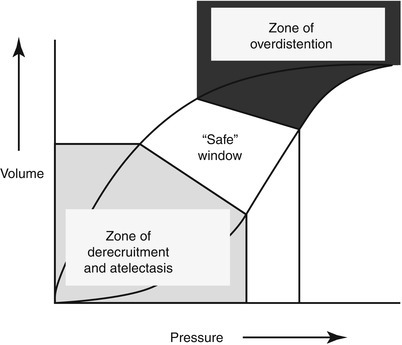
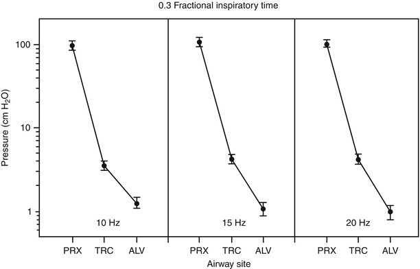
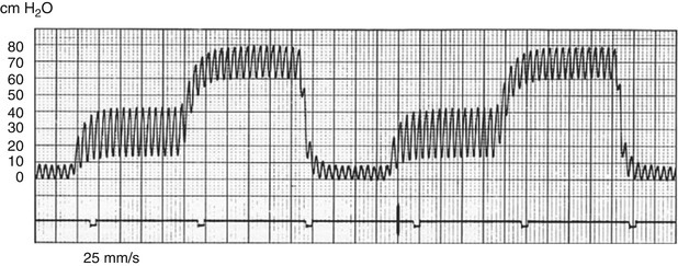
Forty years have elapsed since investigators first appreciated that tidal volumes measuring less than the physiologic dead space can produce reliable ventilation when delivered at high frequencies. Of all high frequency ventilation techniques, high frequency oscillatory ventilation (HFOV) is the most well studied and is the most commonly utilized in clinical practice today. In HFOV, small volume oscillatory vibrations are superimposed on continuous distending pressure in a manner that allows efficient CO2 elimination during continuous alveolar recruitment. By preserving end-expiratory lung volume, minimizing cyclic stretch, and avoiding alveolar overdistension at end-inspiration, HFOV is uniquely capable of providing the ultimate “open lung” strategy of ventilation. Over the past decade, a growing evidence base implicating phasic alveolar stretch in the pathogenesis of acute and chronic lung injury in patients with respiratory failure has driven the iterative refinement of HFOV management protocols for infants, children, and adults. The next step toward applying HFOV in a manner that takes into account the heterogeneity of parenchymal involvement in diseases such as the acute respiratory distress syndrome will require the development of non-invasive bedside technologies capable of identifying regional changes in lung volume and lung mechanics. Electrical impedance tomography (EIT) is a promising technique that could play a supporting role in the conduct of future clinical trials seeking to identify HFOV strategies that are maximally lung protective.


Figures

Fig. 10.1
Fig. 10.1
(ad): Lung volume during conventional ventilation (Panels a, b) compared to HFOV (Panels c, d), at equivalent mean airway pressure. Excised lungs from a rabbit lung lavage model are shown. Panel (a) depicts marked atelectasis at end-expiration (PEEP 9 cm H2O). Panel (b) shows the lung at end inspiration; tidal volume is adjusted to produce eucapnea. Panels (c, d) depict the same lung during HFOV, using a mean airway pressure equivalent to the one represented in Panels (a, b). Panel (c) shows the lung during HFOV without a preceding recruitment maneuver; residual atelectasis remains apparent. Panel (d) shows the lung during HFOV with a preceding recruitment maneuver (Reprinted from Kolton et al. [29]. With permission from Wolter Kluwers Health)
Fig. 10.2
Fig. 10.2
Pressure-volume relationships in acute lung injury. High end-expiratory pressures and small tidal volumes minimize the potential for derecruitment (lower left) and overdistension (upper right). The critical opening pressure of the lung corresponds to the lower inflection point on the inspiratory limb of the volume-pressure curve. The closing pressure of the lung corresponds to the lower inflection point on the expiratory limb of the curve (Reprinted from Froese [129]. With permission from Wolter Kluwers Health)
Fig. 10.3
Fig. 10.3
Amplitude attenuation on HFOV, in open-chested rabbits (without lung injury): The relationship between proximal, tracheal, and alveolar amplitudes are shown at 10, 15, and 20 Hz, and %I:E 0.3 (plotted values represent mean peak-to-trough pressure ± SEM). Proximal pressures are measured at the airway opening. Tracheal pressures are measured 2 cm below the distal opening of the 3.0 mm (outer diameter) endotracheal tube. Alveolar pressures are measured using a pressure transducer attached to a low mass capsule mounted on the pleural surface. Panels depict significant, progressive amplitude attenuation across the endotracheal tube, from airway opening (“PRX”) to trachea (“TRC”) and down to the alveolus (“ALV”) (p < 0.0001) (Reprinted from Gerstmann et al. [35]. With permission from Nature Publishing Group)
Fig. 10.4
Fig. 10.4
Effect of frequency, amplitude, and ETT diameter on tidal volume delivery during HFOV. Data shown in this figure were collected during ventilation of a test lung (MI Instruments, Grand Rapids, MI) with a 3100B oscillator. In these experiments, bias flow is constant at 30 L/min, compliance is constant at 30 mL/cm H2O, and I:E is constant at 1:2. Tidal volume is measured using an adult hot wire anemometer. Panel (a) depicts the relationship between tidal volume and pressure amplitude at a range of frequencies (4–12 Hz), using an 8 mm (inner diameter) endotracheal tube (ETT). Increasing frequency by 2 Hz reduces tidal volume by an average of 21.3 ± 4.1 %. A similar frequency-tidal volume relationship was confirmed by the investigators in a series of adult patients with ARDS, intubated with an 8 mm ETT. In these patients, increasing amplitude by 10 cm H2O produced an average tidal volume increase of only 5.6 ± 4.5 %. Panel (b) depicts the effect of ETT diameter on the relationship between tidal volume and pressure amplitude, at 4 and 12 Hz (Reprinted from Hager et al. [30]. With permission Wolters Kluwer Health)
Fig. 10.5
Fig. 10.5
Transitioning the critically ill child from conventional mechanical ventilatory support to HFOV HFOV initiation, maintenance, and weaning parameters. 1 See text for airleak strategy modifications, 2 Recruitment maneuvers can precipitate acute hemodynamic compromise and should not be routinely performed in patients with hypotension or active airleak. Careful hemodynamic monitoring is advised, and recruitment maneuvers should cease if hypotension occurs [44], 3 Magnitude and extent of chest wall vibrations will vary according to chest wall and/or abdominal compartment compliance, 4 To maximize the lung protective effects of HFOV, the maintenance frequency setting should target the upper limit of each age-based range, 5 Suctioning the poorly compliant lung can result in rapid desaturation. Preoxygenation is recommended, 6 This protocol presumes permissive hypercapnea with target pH ≥ 7.25. This approach is not recommended if there are clear contraindications to permissive hypercapnea (e.g., increased intracranial pressure), 7 Increasing frequency can affect oxygenation by reducing the % inspiratory time. Monitor oxygenation carefully as frequency is adjusted upward (Adapted from Ventre and Arnold [130]. With permission from Elsevier)
Fig. 10.6
Fig. 10.6
Alveolar recruitment along the pressure-volume curve in ARDS: Data shown are from a large animal, oleic acid lung injury model. As lung volume increases toward total lung capacity, aeration of dependent lung units increases substantially, but at a very high airway pressure cost. At high airway pressures, non-dependent lung units may be vulnerable to overdistension. “R” indicates the percentage of total lung recruitment at each corresponding airway pressure (Reprinted with permission of the American Thoracic Society. Copyright (c) 2013 American Thoracic Society. Gattinoni et al. [95]. Official Journal of the American Thoracic Society)
Fig. 10.7
Fig. 10.7
EIT image of the lung. The orientation is the same as for a CT image. Both lung fields show equal impedance change during spontaneous breathing (Adapted from Wolf and Arnold [96]. With permission from Springer Science + Business Media)
Fig. 10.8
Fig. 10.8
Three dimensional depiction of recruitment after suctioning on HFOV. The standard deviation of impedance change after reconnection to the ventilator is displayed (Reprinted from Wolf and Arnold [104]. With permission from Wolter Kluwers Health)
Fig. 10.9
Fig. 10.9
Pressure-time waveform during HFPV (“convective pressure rise” feature engaged): stepwise progression to end inspiratory pressure is depicted. At the beginning of inspiration, oscillatory pressures reach an initial plateau. A “convective pressure rise” carries the breath toward the peak equilibrium pressure, which is then released at the end of inspiration toward the baseline PEEP (CPAP). In this tracing, oscillations are activated during both the inspiratory and the expiratory phase (From The VDR-4 Manual of Understanding [109], used with permission granted by Dr. Forrest Bird)

Comment on

References

    1. Scotter DR, Thurtell GW, Raats PAC. Dispersion resulting from sinusoidal gas flow in porous materials. Soil Sci. 1967;104:306–8. doi: 10.1097/00010694-196710000-00012. - DOI
    1. Bohn DJ, Miyasaka K, Marchak BE, Thompson WK, Froese AB, Bryan AC. Ventilation by high-frequency oscillation. J Appl Physiol. 1980;48(4):710–6. - PubMed
    1. Bryan A. The oscillations of HFO. Am J Respir Crit Care Med. 2001;163:816–7. doi: 10.1164/ajrccm.163.4.16341. - DOI - PubMed
    1. Lunkenheimer PP, Frank I, Ising H, Keller H, Dickhut HH. Intrapulmonary gas exchange during simulated apnea due to transtracheal periodic intrathoracic pressure changes. Anaesthesist. 1973;22(5):232–8. - PubMed
    1. Lunkenheimer PP, Rafflenbeul W, Keller H, Frank I, Dickhut HH, Fuhrmann C. Application of transtracheal pressure oscillations as a modification of “diffusing respiration”. Br J Anaesth. 1972;44(6):627. doi: 10.1093/bja/44.6.627. - DOI - PubMed

MeSH terms