Review
. 2014 Apr:5:16-23.
doi: 10.1016/j.coviro.2014.01.004. Epub 2014 Feb 1.

Structural basis of efficient contagion: measles variations on a theme by parainfluenza viruses

Affiliations
Review

Structural basis of efficient contagion: measles variations on a theme by parainfluenza viruses

Mathieu Mateo et al. Curr Opin Virol. 2014 Apr.

Abstract

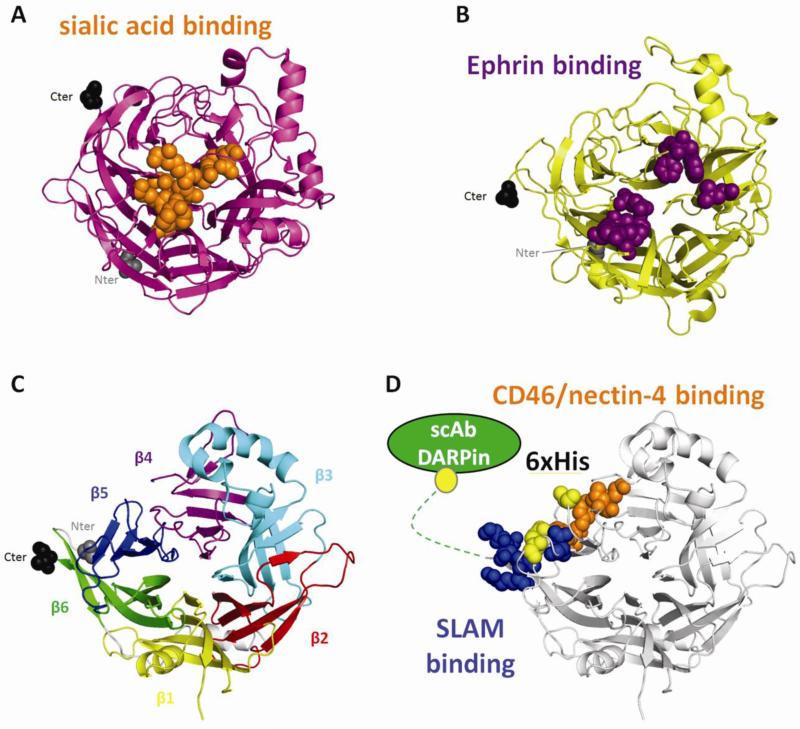
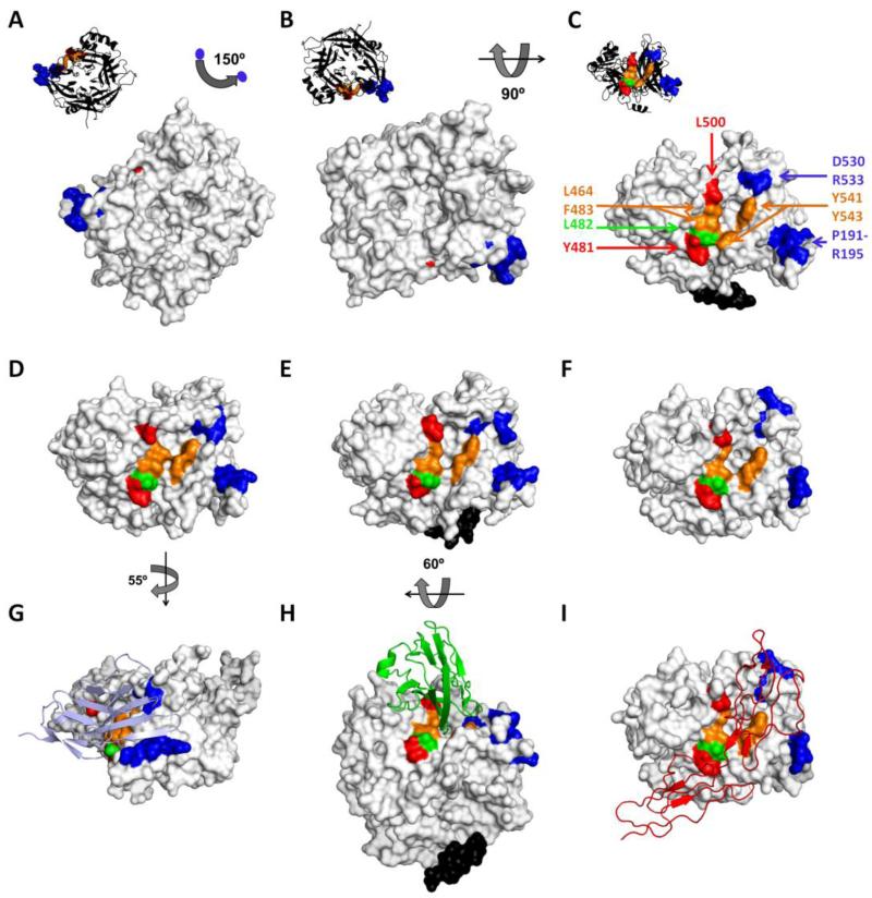
A quartet of attachment proteins and a trio of fusion protein subunits play the cell entry concert of parainfluenza viruses. While many of these viruses bind sialic acid to enter cells, wild type measles binds exclusively two tissue-specific proteins, the lymphatic receptor signaling lymphocytic activation molecule (SLAM), and the epithelial receptor nectin-4. SLAM binds near the stalk-head junction of the hemagglutinin. Nectin-4 binds a hydrophobic groove located between blades 4 and 5 of the hemagglutinin β-propeller head. The mutated vaccine strain hemagglutinin binds in addition the ubiquitous protein CD46, which explains attenuation. The measles virus entry concert has four movements. Andante misterioso: the virus takes over the immune system. Allegro con brio: it rapidly spreads in the upper airway's epithelia. 'Targeting' fugue: the versatile orchestra takes off. Presto furioso: the virus exits the host with thunder. Be careful: music is contagious.


Figures

Figure 1
Figure 1
Structures of paramyxovirus attachement protein heads and modes of receptor binding. (A) Structure of PIV5 HN. Residues contacting the sialic acid are shown in orange. (B) Structure of Nipah G. Residues contacting ephrin B2 are colored purple. (C) Structure of MeV H. Each blade in the 6-bladed β-propeller head is represented in a different color. (D) MeV H showing the most important residues binding CD46/nectin-4 (orange), those most important for binding SLAM (blue), the positions of hexahistidine tags supporting fusion (yellow), and C-terminal addition of specificity domains (green). In A, B and C, the N-terminal residue and the C-terminal residues are in spherical representation grey and black, respectively. The structures were aligned using PyMOL (www.pymol.org).
Figure 2
Figure 2
Surface representation of the MeV H head unbound (A-C) [11], after SLAM binding (D) [50], after nectin-4 binding (E) [54] or after CD46 binding (F) [58]. With the exception of the H-nectin-4 co-crystal obtained with a wild-type H protein, all other structures were based on vaccine-lineage H proteins. In panel A, MV H has the same orientation as in Figure 1C, as shown also in the upper left black ribbon backbone representation, and is rotated according to the indicated angles in B and C. D, E and F panels are oriented as in C. G, H and I show also the receptors in ribbon representation: SLAM (light blue), nectin-4 (green) and CD46 (red), respectively. The MeV H residues important for function through CD46, nectin-4 and SLAM are colored red, green and blue, respectively. Residues shared by the CD46 and nectin-4 binding sites are colored orange. Stalk-connecting residues are colored black. Certain parts of the H protein structures were not resolved, also accounting for part of the visible differences between the structures. For example, the upper stalk residues 156 to 166 are only visible in the H/nectin-4 and unbound structures (panels C, E and H, black residues). Minor differences in structure noted upon binding to different receptors are mainly due to the orientation of the amino acid side chains (see main text).
Figure 3
Figure 3
Two possible poses of the MV H-head dimers on the stalk. A) The H-head dimers are depicted in a 4-heads-down orientation similar to that seen for HN [14]. One monomer in each H-dimer is colored such that each β-propeller blade is shaded differently, as in Fig. 1A. The other monomer is shaded grey. Cysteine (Cys) 154 at the top of the stalk is shown in a space-filling representation and shaded yellow. A large gap in the crystal structure which corresponds to a flexible loop in the head-connecting segment is shown as a purple wavy line. Each horizontal line represents a residue in the stalk which was systematically substituted with Cys [73]. Numbering between the panels refers to MV H-stalk residues. Length of the horizontal line is proportional to the covalent tetramer trapping propensity of the Cys substitution at that position. The color of the line indicates the fusion support efficiency of the Cys substitutions: green, wild type levels of fusion; blue, intermediate fusion; orange, greatly reduced fusion; and red, no fusion. B) The H-head dimers are depicted in the form I arrangement [50]. Since a mutated H-head construct lacking the head-connecting segment was used to crystallize the form I tetramer, the head connecting segment of the original H-head dimer structure [11] was modeled into the form I structure. While modeling revealed some steric clash issues, all four Cys154 residues come together at the top of the stalk in this H-head conformation.

References

    1. Griffin D. Measles. In: Knipe D, Howley P, editors. Fields’ Virology. Sixth Edition Vol. 1. Lippincott, Williams and Wilkins; 2013. pp. 1042–1069.
    1. Lamb RA, Parks G. Paramyxoviridae. In: Knipe D, Howley P, editors. Fields’ Virology. Sixth edition Vol. 1. Lippincott, Williams & Wilkins; 2013. pp. 957–995.
    1. Lee B, Ataman ZA. Modes of paramyxovirus fusion: a Henipavirus perspective. Trends Microbiol. 2011;19:389–399. - PMC - PubMed
    1. Plattet P, Plemper RK. Envelope protein dynamics in paramyxovirus entry. MBio. 2013;4:e00413–13. - PMC - PubMed
    1. Modis Y. Relating structure to evolution in class II viral membrane fusion proteins. Curr Opin Virol. 2014 COVIRO-S-13-00089. - PMC - PubMed

Publication types

MeSH terms