

Evaluation of human pancreatic RNase as effector molecule in a therapeutic antibody platform
- PMID: 24492302
- PMCID: PMC3984326
- DOI: 10.4161/mabs.27830
Evaluation of human pancreatic RNase as effector molecule in a therapeutic antibody platform
Abstract
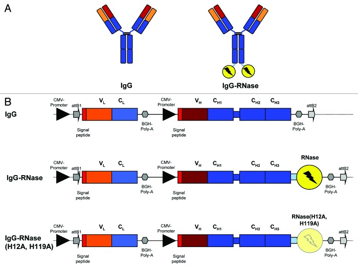
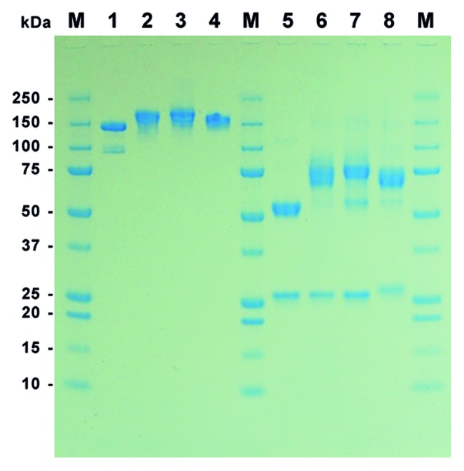
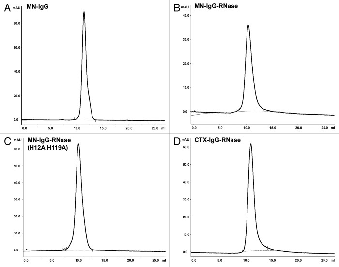
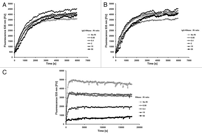
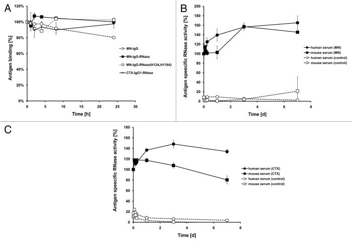
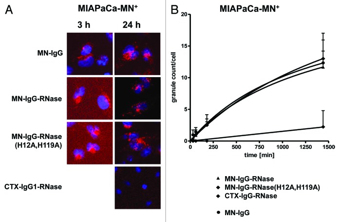
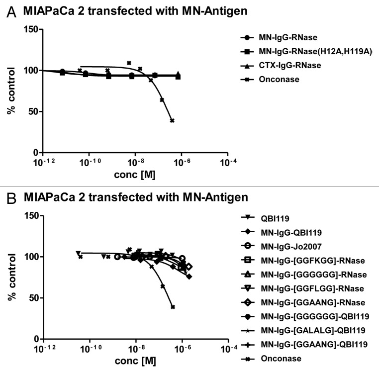
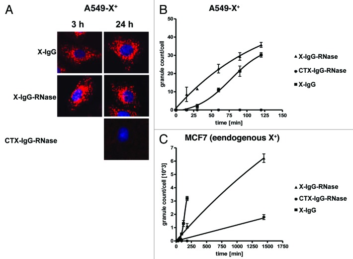
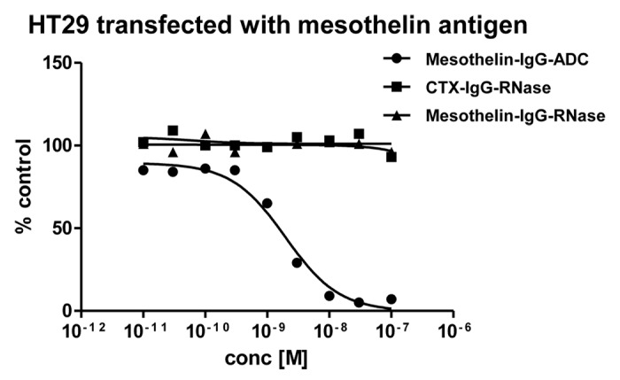
Human antibody-ribonuclease (RNase) fusion proteins, referred to as immunoRNases, have been proposed as an alternative to heterologous immunotoxins, without their immunogenicity and unspecific toxicity issues. In this study, we investigated if human pancreatic RNase will be suitable as effector component in a therapeutic antibody development platform. We generated several fusion proteins consisting of tumor-specific human immunoglobulins (IgGs) and human pancreatic RNase. Transient mammalian cell production was efficient and IgG-RNases were purified to homogeneity. Antigen binding was comparable to the parental antibodies and RNase catalytic activity was retained even in the presence of 50-fold molar excess of human cytosolic RNase inhibitor (RI). Serum stability, cell binding and internalization of IgG-RNases were comparable to the parental IgGs. Despite these promising properties, none of the IgG-RNases revealed significant inhibition of tumor cell growth in vitro even when targeting different antigens putatively employing different endocytotic pathways. The introduction of different linkers containing endosomal protease cleavage sites into the IgG-RNase did not enhance cytotoxicity. Similarly, RI evasive human pancreatic RNase variants mediated only small inhibiting effects on tumor cell growth at high concentrations, potentially reflecting inefficient cytosolic translocation. Taken together, human pancreatic RNase and variants did not prove to be generally suitable as effector component for a therapeutic antibody drug development platform.
Keywords: IgG; RNase inhibitor; antibodies; cancer therapy; human pancreatic RNase; immunoRNase; immunoglobulin.
Figures
Similar articles
-
Antibody fusion proteins with human ribonucleases 1 to 8.Hum Antibodies. 2018;26(4):177-192. doi: 10.3233/HAB-180337. Hum Antibodies. 2018. PMID: 29689715
-
Endocytotic internalization as a crucial factor for the cytotoxicity of ribonucleases.J Biol Chem. 2007 Sep 21;282(38):27640-6. doi: 10.1074/jbc.M702240200. Epub 2007 Jul 17. J Biol Chem. 2007. PMID: 17635931
-
A novel fully human antitumor immunoRNase resistant to the RNase inhibitor.Protein Eng Des Sel. 2013 Mar;26(3):243-8. doi: 10.1093/protein/gzs101. Epub 2012 Dec 11. Protein Eng Des Sel. 2013. PMID: 23232187 Free PMC article.
-
Refined immunoRNases for the efficient targeting and selective killing of tumour cells: A novel strategy.Life Sci. 2022 Jan 15;289:120222. doi: 10.1016/j.lfs.2021.120222. Epub 2021 Dec 10. Life Sci. 2022. PMID: 34902436 Review.
-
From immunotoxins to immunoRNases.Curr Pharm Biotechnol. 2008 Jun;9(3):210-4. doi: 10.2174/138920108784567254. Curr Pharm Biotechnol. 2008. PMID: 18673286 Review.
Cited by
-
Endosomal escape efficiency of fusogenic B18 and B55 peptides fused with anti-EGFR single chain Fv as estimated by nuclear translocation.J Biochem. 2016 Jan;159(1):123-32. doi: 10.1093/jb/mvv083. Epub 2015 Sep 2. J Biochem. 2016. PMID: 26338729 Free PMC article.
-
A novel Carcinoembryonic Antigen (CEA)-Targeted Trimeric Immunotoxin shows significantly enhanced Antitumor Activity in Human Colorectal Cancer Xenografts.Sci Rep. 2019 Aug 12;9(1):11680. doi: 10.1038/s41598-019-48285-z. Sci Rep. 2019. PMID: 31406218 Free PMC article.
-
Updates in the Development of ImmunoRNases for the Selective Killing of Tumor Cells.Biomedicines. 2018 Mar 5;6(1):28. doi: 10.3390/biomedicines6010028. Biomedicines. 2018. PMID: 29510557 Free PMC article. Review.
-
Inclusion of a Furin Cleavage Site Enhances Antitumor Efficacy against Colorectal Cancer Cells of Ribotoxin α-Sarcin- or RNase T1-Based Immunotoxins.Toxins (Basel). 2019 Oct 12;11(10):593. doi: 10.3390/toxins11100593. Toxins (Basel). 2019. PMID: 31614771 Free PMC article.
-
Addressing the Immunogenicity of the Cargo and of the Targeting Antibodies with a Focus on Demmunized Bacterial Toxins and on Antibody-Targeted Human Effector Proteins.Biomedicines. 2017 Jun 2;5(2):28. doi: 10.3390/biomedicines5020028. Biomedicines. 2017. PMID: 28574434 Free PMC article. Review.
References
-
- Muirhead M, Martin PJ, Torok-Storb B, Uhr JW, Vitetta ES. Use of an antibody-ricin A-chain conjugate to delete neoplastic B cells from human bone marrow. Blood. 1983;62:327–32. - PubMed
-
- Giles FJ, Kantarjian HM, Kornblau SM, Thomas DA, Garcia-Manero G, Waddelow TA, David CL, Phan AT, Colburn DE, Rashid A, et al. Mylotarg (gemtuzumab ozogamicin) therapy is associated with hepatic venoocclusive disease in patients who have not received stem cell transplantation. Cancer. 2001;92:406–13. doi: 10.1002/1097-0142(20010715)92:2<406::AID-CNCR1336>3.0.CO;2-U. - DOI - PubMed
MeSH terms
Substances
LinkOut - more resources
Full Text Sources
Other Literature Sources
Medical