. 2014 Feb 18;111(7):2686-91.
doi: 10.1073/pnas.1400419111. Epub 2014 Feb 3.

Blocking CD40-TRAF6 signaling is a therapeutic target in obesity-associated insulin resistance

Affiliations

Blocking CD40-TRAF6 signaling is a therapeutic target in obesity-associated insulin resistance

Antonios Chatzigeorgiou et al. Proc Natl Acad Sci U S A. .

Erratum in

  • Proc Natl Acad Sci U S A. 2014 Mar 25;111(12):4644. Schulte, Klaus [corrected to Schulte, Klaus-Martin]

Abstract

The immune system plays an instrumental role in obesity and insulin resistance. Here, we unravel the role of the costimulatory molecule CD40 and its signaling intermediates, TNF receptor-associated factors (TRAFs), in diet-induced obesity (DIO). Although not exhibiting increased weight gain, male CD40(-/-) mice in DIO displayed worsened insulin resistance, compared with wild-type mice. This worsening was associated with excessive inflammation of adipose tissue (AT), characterized by increased accumulation of CD8(+) T cells and M1 macrophages, and enhanced hepatosteatosis. Mice with deficient CD40-TRAF2/3/5 signaling in MHCII(+) cells exhibited a similar phenotype in DIO as CD40(-/-) mice. In contrast, mice with deficient CD40-TRAF6 signaling in MHCII(+) cells displayed no insulin resistance and showed a reduction in both AT inflammation and hepatosteatosis in DIO. To prove the therapeutic potential of inhibition of CD40-TRAF6 in obesity, DIO mice were treated with a small-molecule inhibitor that we designed to specifically block CD40-TRAF6 interactions; this compound improved insulin sensitivity, reduced AT inflammation, and decreased hepatosteatosis. Our study reveals that the CD40-TRAF2/3/5 signaling pathway in MHCII(+) cells protects against AT inflammation and metabolic complications associated with obesity whereas CD40-TRAF6 interactions in MHCII(+) cells aggravate these complications. Inhibition of CD40-TRAF6 signaling by our compound may provide a therapeutic option in obesity-associated insulin resistance.

Keywords: immunity; metabolism; type 2 diabetes.


Conflict of interest statement

The authors declare no conflict of interest.

Figures
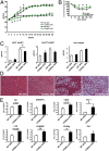
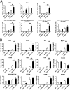
Fig. 1.
Fig. 1.
CD40−/− mice on HFD display aggravated metabolic dysregulation. Wild-type (WT) and CD40−/− male mice were fed with SFD or HFD for 30 wk. (A) Body weight of WT and CD40−/− mice on SFD (n = 5) or HFD (n = 6–7). (B) Insulin tolerance test (ITT) in 5-h fasted WT and CD40−/− mice fed an HFD for 30 wk (n = 6–7). (C) Weights of sqAT, gonAT, and liver of WT and CD40−/− mice after 30 wk on SFD (n = 5) or HFD (n = 6–7). (D) Representative H&E-stained sections from liver of WT and CD40−/− mice on SFD or HFD for 30 wk. (E) Liver gene expression of WT and CD40−/− mice on HFD for 30 wk. The mRNA expression was normalized against 18S, and the gene expression of livers from WT HFD was set as 1 (n = 6–7). *P < 0.05 for comparison between WT and CD40−/− mice fed the same diet.
Fig. 2.
Fig. 2.
CD40 deficiency aggravates AT inflammation. (A) SVF cells were isolated from gonAT of WT or CD40−/− male mice fed an SFD or HFD for 18 wk. CD45+ leukocytes, CD4+ or CD8+ lymphocytes, total macrophages (characterized as CD11b+F4/80+), and M1 macrophages (defined as F4/80+CD11b+CD11c+ or F4/80+CD11c+CD206) or M2 macrophages (defined as F4/80+CD11cCD206+) were analyzed by flow cytometry. (B) Gene expression in the gonAT of male WT and CD40−/− mice on SFD or HFD for 18 wk. The mRNA expression was normalized against 18S, and the gene expression of adipose tissue from WT SFD was set as 1. n = 4 or more per group for SFD groups and n = 7 or more per group for HFD groups. *P < 0.05 for comparison between WT and CD40−/− mice fed the same diet.
Fig. 3.
Fig. 3.
Deficiency of CD40-TRAF2/3/5 signaling aggravates obesity and promotes metabolic dysfunction and hepatosteatosis. (A) Body weight of CD40-Twt, CD40-T2/3/5−/−, and CD40-T6−/− mice on SFD or HFD for 20 wk (n = 12–15). (B) Fat mass as determined by 1H NMR spectroscopy (n = 8 per group). (C) ITT in 5-h fasted CD40-Twt, CD40-T2/3/5−/−, and CD40-T6−/− mice fed an HFD for 18 wk (n = 8 per group). (D) Liver weight and (E) Oil red O-stained liver cryosections of CD40-Twt, CD40-T2/3/5−/−, and CD40-T6−/− mice fed an HFD for 20 wk. (Scale bar: 100 μm.) Values are mean ± SEM. *P < 0.05 for comparison with CD40-Twt mice.
Fig. 4.
Fig. 4.
CD40-TRAF2/3/5 deficiency results in increased numbers of inflammatory cells in gonadal AT in DIO. (A) FACS analysis for F4/80highCD11b+ macrophages, (B) CD3+ T cells, CD8+ T cells, and CD4+ T cells, (C) CD4+CD25+FoxP3+ Tregs, and (D) CD8+CD44lowCD62Lhigh naïve T cells and CD8+CD44highCD62Llow effector T cells. Values are mean ± SEM. *P < 0.05 for comparison with CD40-Twt mice. n = 8–10 mice per group.
Fig. 5.
Fig. 5.
CD40-TRAF6 interaction inhibitor improves metabolic dysregulation and AT inflammation. WT male mice were fed an HFD for a total of 12 wk, receiving a CD40-TRAF6 interaction inhibitor (6877002) (10 μmol⋅kg−1⋅d−1 i.p.) or vehicle starting at week 6 of feeding. (A) Body weight of mice fed an HFD for 12 wk and treated with inhibitor or control. (B) ITT of mice fed an HFD for 12 wk and treated with inhibitor or control. (C) SVF cells from gonAT of control- or inhibitor-treated mice were analyzed by FACS. CD45+ leukocytes and total macrophages or M1 macrophages, characterized as CD11b+F4/80+ and F4/80+CD11b+CD11c+, respectively, are shown. (D) Representative H&E-stained sections from livers of control- or inhibitor-treated mice. *P < 0.05 for comparison with control-treated mice; n = 7–8 mice per group.

References

    1. Osborn O, Sears DD, Olefsky JM. Fat-induced inflammation unchecked. Cell Metab. 2010;12(6):553–554. - PubMed
    1. Chatzigeorgiou A, Karalis KP, Bornstein SR, Chavakis T. Lymphocytes in obesity-related adipose tissue inflammation. Diabetologia. 2012;55(10):2583–2592. - PubMed
    1. Gregor MF, Hotamisligil GS. Inflammatory mechanisms in obesity. Annu Rev Immunol. 2011;29:415–445. - PubMed
    1. Nishimura S, et al. CD8+ effector T cells contribute to macrophage recruitment and adipose tissue inflammation in obesity. Nat Med. 2009;15(8):914–920. - PubMed
    1. Feuerer M, et al. Lean, but not obese, fat is enriched for a unique population of regulatory T cells that affect metabolic parameters. Nat Med. 2009;15(8):930–939. - PMC - PubMed

Publication types

MeSH terms