. 2014 Apr 15;592(8):1823-38.
doi: 10.1113/jphysiol.2013.268086. Epub 2014 Feb 3.

GABAergic and glycinergic inputs modulate rhythmogenic mechanisms in the lamprey respiratory network

Affiliations

GABAergic and glycinergic inputs modulate rhythmogenic mechanisms in the lamprey respiratory network

Elenia Cinelli et al. J Physiol. .

Abstract

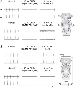
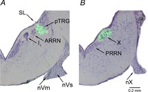
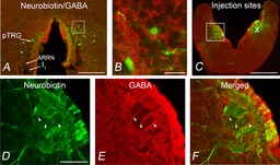
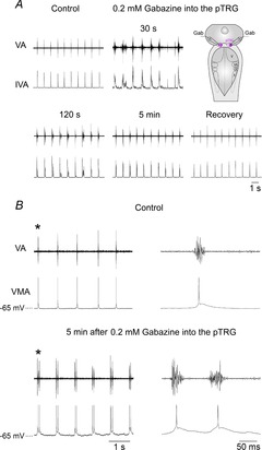
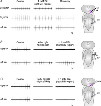
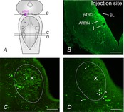
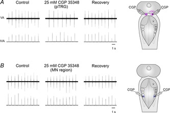
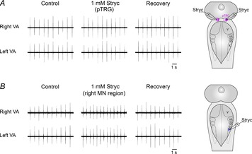
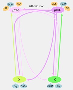
We have previously shown that GABA and glycine modulate respiratory activity in the in vitro brainstem preparations of the lamprey and that blockade of GABAA and glycine receptors restores the respiratory rhythm during apnoea caused by blockade of ionotropic glutamate receptors. However, the neural substrates involved in these effects are unknown. To address this issue, the role of GABAA, GABAB and glycine receptors within the paratrigeminal respiratory group (pTRG), the proposed respiratory central pattern generator, and the vagal motoneuron region was investigated both during apnoea induced by blockade of glutamatergic transmission and under basal conditions through microinjections of specific antagonists. The removal of GABAergic, but not glycinergic transmission within the pTRG, causes the resumption of rhythmic respiratory activity during apnoea, and reveals the presence of a modulatory control of the pTRG under basal conditions. A blockade of GABAA and glycine receptors within the vagal region strongly increases the respiratory frequency through disinhibition of neurons projecting to the pTRG from the vagal region. These neurons were retrogradely labelled (neurobiotin) from the pTRG. Intense GABA immunoreactivity is observed both within the pTRG and the vagal area, which corroborates present findings. The results confirm the pTRG as a primary site of respiratory rhythm generation, and suggest that inhibition modulates the activity of rhythm-generating neurons, without any direct role in burst formation and termination mechanisms.


Figures

Figure 1
Figure 1. Role of GABAA receptors in the resumption of respiratory activity during blockade of ionotropic glutamate receptors
A, in one preparation, during apnoea caused by bath application of 20 μm CNQX and 100 μm d-AP5, bilateral microinjections of 1 mm bicuculline (Bic) into the pTRG restored the respiratory rhythm within 4 min after the completion of the injections, while similar microinjections into the region of vagal motoneurons (MNs) caused the appearance of a low level of tonic vagal activity. The sites (pink area, pTRG; blue area, vagal MN region) where Bic was microinjected are shown on a schematic illustration of a dorsal view of the lamprey mesencephalon–rhombencephalon. B, in another preparation, bath application of 20 μm CNQX and 100 μm d-AP5 abolished the respiratory rhythm that was restored by 10 μm Bic, but not by 10 μm strychnine (Stryc). A schematic illustration of a dorsal view of the lamprey mesencephalon–rhombencephalon in the perfused recording chamber is shown. pTRG, paratrigeminal respiratory group; V, trigeminal motor nucleus; VII, facial motor nucleus; IX, glossopharyngeal motor nucleus; X, vagal motor nucleus; VA, raw vagal nerve activity; IVA, integrated vagal nerve activity.
Figure 2
Figure 2. Localization of injection sites
Photomicrographs of transverse sections of the rhombencephalon showing the location of the fluorescent beads (green) added to 1 mm bicuculline microinjected into the pTRG (A) and the vagal motor nucleus (B). Sections are counterstained with Cresyl Violet. Light-field and fluorescence photomicrographs have been superimposed. ARRN, anterior rhombencephalic reticular nucleus; I1, isthmic Müller cell; nVm, motor root of the trigeminal nerve; nVs, sensory root of the trigeminal nerve; nX, vagal nerve; PRRN, posterior rhombencephalic reticular nucleus; pTRG, paratrigeminal respiratory group; SL, sulcus limitans of His; X, vagal motor nucleus.
Figure 3
Figure 3. Distribution of GABA immunoreactivity in the pTRG region and the vagal motor nucleus
A, photomicrograph of a transverse section from the isthmic region at the level of the pTRG showing retrogradely labelled neurons (green) after bilateral injections of neurobiotin into the vagal motoneuron pools. GABA immunoreactivity is shown in red. B, photomicrograph at a higher magnification of the portion of the transverse section (white box in A) showing retrogradely labelled neurons in the pTRG surrounded by GABA-immunoreactive structures. C, photomicrograph of a transverse section in the caudal rhombencephalon showing the site of bilateral neurobiotin injections into the vagal motor nuclei. The distribution of GABA immunoreactivity is shown in red. D–F, photomicrographs at a higher magnification of the portion of the transverse section indicated by the white box in C. Arrowheads point to GABAergic fibres surrounding the cell bodies of vagal motoneurons. D, vagal motoneurons stained by neurobiotin injection. E, GABA-immunoreactive structures. F, merged image. Scale bars, 200 μm in A, 25 μm in B, 500 μm in C, 100 μm in D–F. ARRN, anterior rhombencephalic reticular nucleus; I1, isthmic Müller cell; pTRG, paratrigeminal respiratory group; X, vagal motor nucleus.
Figure 4
Figure 4. Respiratory role of GABAA receptors within the pTRG in one brainstem preparation
A, respiratory responses induced by bilateral microinjections of 0.2 mm gabazine into the pTRG at different time points after the completion of the injections. Traces are raw vagal nerve activity (VA) and integrated vagal nerve activity (IVA). The microinjections caused within 20–30 s the appearance of irregular, prolonged vagal bursts (seizure-like activity) associated with tonic activity. Thereafter, a respiratory activity characterized by rhythmic bursts assembled in multiple bursts developed progressively and stabilized in a double-burst pattern. The sites where gabazine (Gab) was microinjected into the pTRG (pink area) are shown on a schematic illustration of a dorsal view of the lamprey mesencephalon–rhombencephalon. B, bilateral microinjections of 0.2 mm gabazine into the pTRG caused the appearance of vagal bursts grouped in duplets within 5 min of the completion of the injections. Traces are raw vagal nerve activity (VA) and vagal motoneuron activity (VMA). The first single or double vagal and neuronal bursts (marked with an asterisk) are shown with an expanded time scale on the right of each trace. Note delayed repolarization in motoneuron membrane potential after gabazine. pTRG, paratrigeminal respiratory group; V, trigeminal motor nucleus; VII, facial motor nucleus; IX, glossopharyngeal motor nucleus; X, vagal motor nucleus.
Figure 5
Figure 5. Respiratory role of GABAA receptors within the vagal motor nucleus
A, marked increases in respiratory frequency ∼4 min after a unilateral microinjection of 1 mm bicuculline (Bic) into the region of vagal motoneurons (MN region) in one preparation. Note the excitatory effects on pTRG neuronal activity (pauci-unit recordings; NA) as well as on the right and left raw vagal nerve activity (VA). Sites where extracellular recordings of pTRG neurons (pink area) and Bic microinjections (blue area) were performed are shown on a schematic illustration of a dorsal view of the lamprey mesencephalon–rhombencephalon. B, in another preparation, hemisection of the brainstem between the pTRG and the vagal motoneuron region abolished the ipsilateral vagal output and slightly reduced the contralateral vagal bursts. Unilateral microinjection of 1 mm Bic into the region of vagal motoneurons on the hemisected side caused slight tonic activity in the ipsilateral vagal output, but did not change the pattern of rhythmic activity on the contralateral side. A schematic illustration of a dorsal view of the lamprey mesencephalon–rhombencephalon showing the hemisection (scissors) and the Bic microinjection site (blue area) is reported. C, during apnoea caused by 1 mm CNQX unilateral microinjection into the pTRG, unilateral microinjection of 1 mm Bic into the region of vagal motoneurons of the same side caused the appearance of a low level of tonic vagal activity on the ipsilateral side of another preparation. Injection sites (pink area, pTRG; blue area, vagal MN region) are schematically depicted on a dorsal view of the lamprey mesencephalon–rhombencephalon. pTRG, paratrigeminal respiratory group; V, trigeminal motor nucleus; VII, facial motor nucleus; IX, glossopharyngeal motor nucleus; X, vagal motor nucleus.
Figure 6
Figure 6. Retrogradely labelled neurons in the vagal motoneuron region
A, schematic illustration of a dorsal view of the lamprey mesencephalon–rhombencephalon showing the levels of the coronal sections illustrated in B–D (continuous lines) and the location of the pTRG (pink area). B, photomicrograph of a transverse section of the isthmic region showing the site of neurobiotin injection into the pTRG (green signal). C and D, photomicrographs of transverse sections, 100 μm apart, of the vagal motoneuron region (dotted line) showing retrogradely labelled neurons (green signal) after an injection of neurobiotin into the pTRG. Arrowheads point to some retrogradely labelled neurons. ARRN, anterior rhombencephalic reticular nucleus; I1, isthmic Müller cell; pTRG, paratrigeminal respiratory group; SL, sulcus limitans of His; V, trigeminal motor nucleus; VII, facial motor nucleus; IX, glossopharyngeal motor nucleus; X, vagal motor nucleus. Scale bars, 200 μm in B, 100 μm in C and D.
Figure 7
Figure 7. Respiratory role of GABAB receptors
A, decreases in respiratory frequency ∼15 min after bilateral microinjections of 25 mm CGP 35348 into the pTRG in one preparation. B, absence of appreciable respiratory effects ∼15 min after bilateral microinjections of 25 mm CGP 35348 into the region of vagal motoneurons (MN region) in a different preparation. The sites (pink area, pTRG; blue area, vagal MN region) where CGP 35348 (CGP) was microinjected are shown on a schematic illustration of a dorsal view of the lamprey mesencephalon–rhombencephalon. pTRG, paratrigeminal respiratory group; V, trigeminal motor nucleus; VII, facial motor nucleus; IX, glossopharyngeal motor nucleus; X, vagal motor nucleus; VA, raw vagal nerve activity; IVA, integrated vagal nerve activity.
Figure 8
Figure 8. Respiratory role of glycine receptors
A, absence of appreciable respiratory effects ∼3 min after bilateral microinjections of 1 mm strychnine (Stryc) into the pTRG in one preparation. B, marked increases in respiratory frequency ∼3 min after a unilateral microinjection of 1 mm Stryc into the region of vagal motoneurons (MN region) in a different preparation. The sites (pink area, pTRG; blue area, vagal MN region) where Stryc was microinjected are shown on a schematic illustration of a dorsal view of the lamprey mesencephalon–rhombencephalon. pTRG, paratrigeminal respiratory group; V, trigeminal motor nucleus; VII, facial motor nucleus; IX, glossopharyngeal motor nucleus; X, vagal motor nucleus; VA, raw vagal nerve activity; IVA, integrated vagal nerve activity.
Figure 9
Figure 9. Schematic drawing representing present and previous findings on the connectivity within the respiratory network and relevant neurotransmitter influences
The pTRG region is shown with its projections (pink) to ipsilateral and contralateral vagal motoneuron regions and to the contralateral pTRG (Gariépy et al. ; Cinelli et al. 2013). Excitatory (yellow) and inhibitory (blue) influences on the pTRG region (present findings and Mutolo et al. , ; Cinelli et al. 2013) and the vagal motoneuron region (present findings) are illustrated. Newly identified excitatory projections to the pTRG (green) from neurons located in the vagal area have also been reported. ACh, acetylcholine; GABA, γ-aminobutyric acid; Gly, glycine; pTRG, paratrigeminal respiratory group region; SP, substance P; X, vagal motoneuron region.

Comment in

References

    1. Bailey TW, Appleyard SM, Jin YH, Andresen MC. Organization and properties of GABAergic neurons in solitary tract nucleus (NTS) J Neurophysiol. 2008;99:1712–1722. - PubMed
    1. Bettler B, Kaupmann K, Mosbacher J, Gassmann M. Molecular structure and physiological functions of GABAB receptors. Physiol Rev. 2004;84:835–867. - PubMed
    1. Bongianni F, Deliagina TG, Grillner S. Role of glutamate receptor subtypes in the lamprey respiratory network. Brain Res. 1999;826:298–302. - PubMed
    1. Bongianni F, Mutolo D, Carfi M, Pantaleo T. Group I and II metabotropic glutamate receptors modulate respiratory activity in the lamprey. Eur J Neurosci. 2002;16:454–460. - PubMed
    1. Bongianni F, Mutolo D, Cinelli E, Pantaleo T. Respiratory responses induced by blockades of GABA and glycine receptors within the Botzinger complex and the pre-Botzinger complex of the rabbit. Brain Res. 2010;1344:134–147. - PubMed

Publication types

MeSH terms

LinkOut - more resources