. 2014 Jan 28;12(1):e1001777.
doi: 10.1371/journal.pbio.1001777. eCollection 2014 Jan.

Drosophila Tempura, a novel protein prenyltransferase α subunit, regulates notch signaling via Rab1 and Rab11

Affiliations

Drosophila Tempura, a novel protein prenyltransferase α subunit, regulates notch signaling via Rab1 and Rab11

Wu-Lin Charng et al. PLoS Biol. .

Abstract

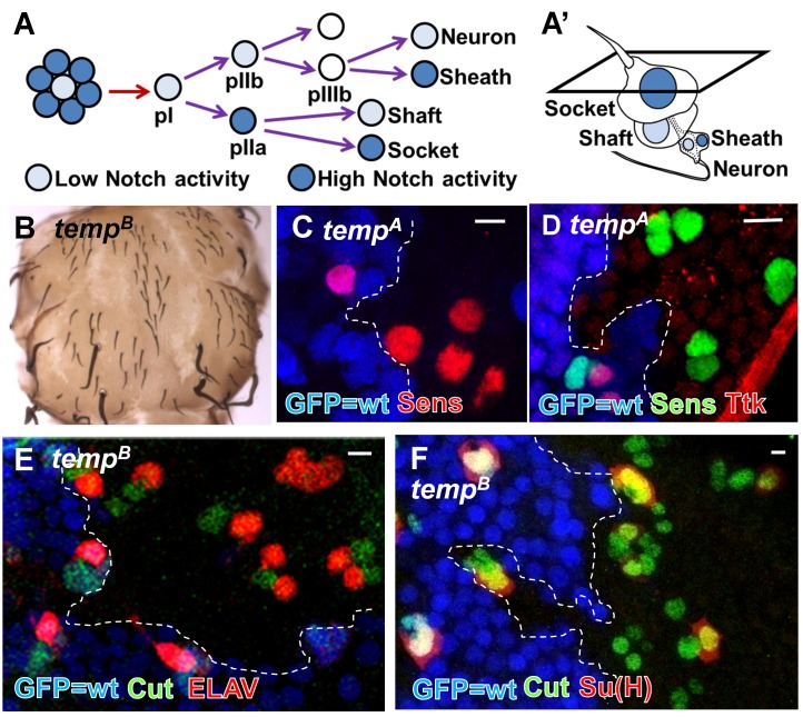
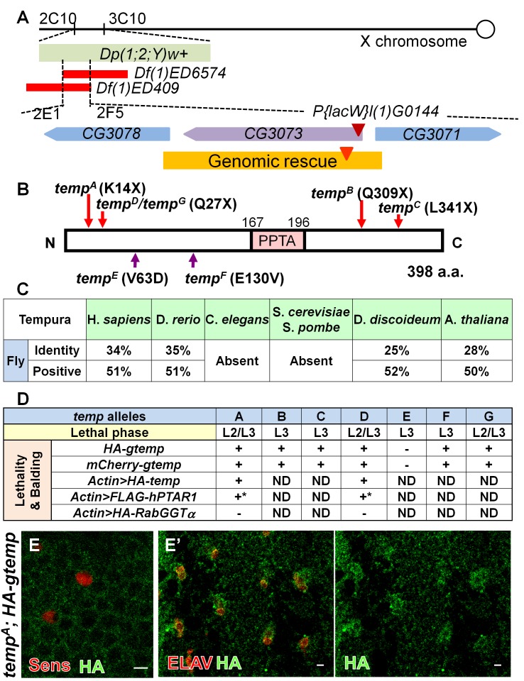
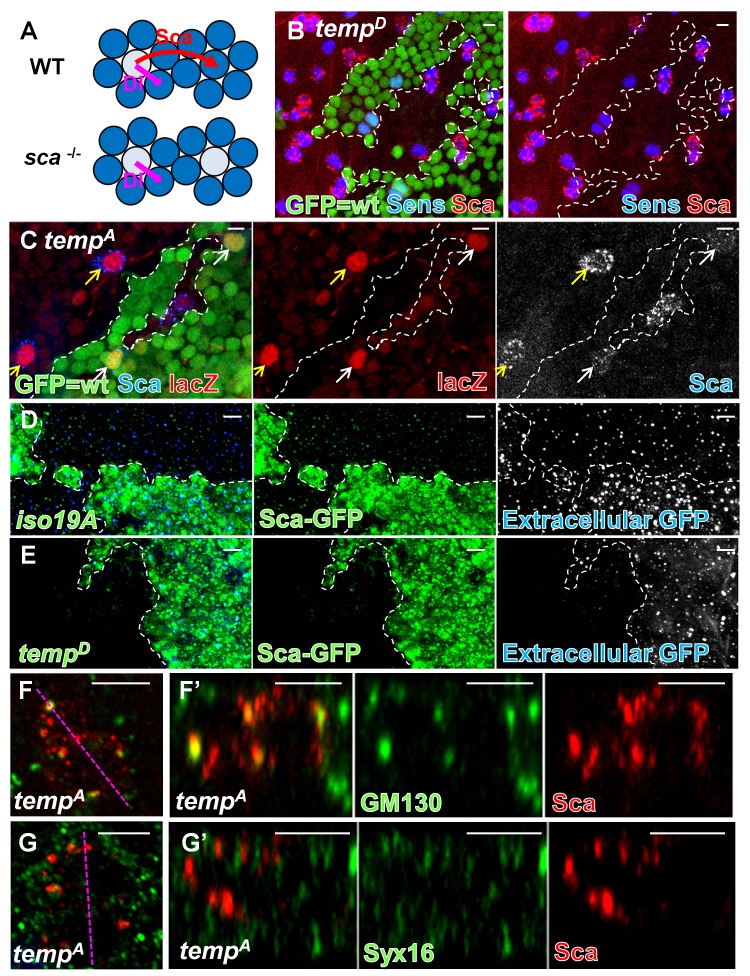
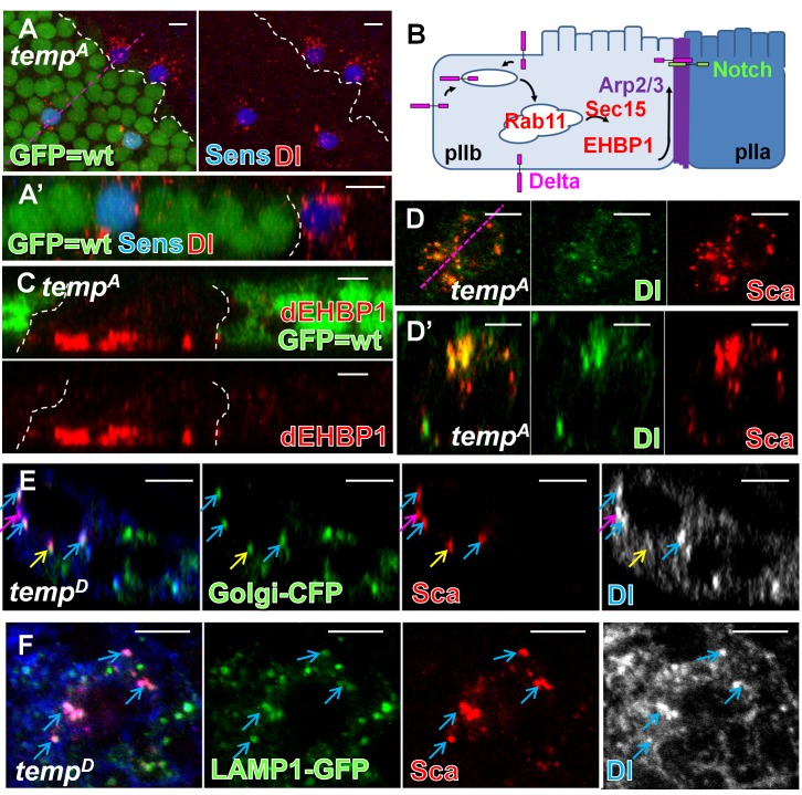
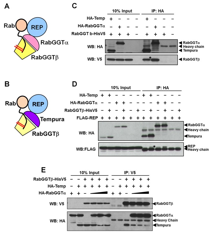
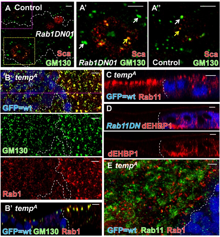
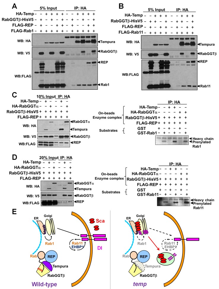
Vesicular trafficking plays a key role in tuning the activity of Notch signaling. Here, we describe a novel and conserved Rab geranylgeranyltransferase (RabGGT)-α-like subunit that is required for Notch signaling-mediated lateral inhibition and cell fate determination of external sensory organs. This protein is encoded by tempura, and its loss affects the secretion of Scabrous and Delta, two proteins required for proper Notch signaling. We show that Tempura forms a heretofore uncharacterized RabGGT complex that geranylgeranylates Rab1 and Rab11. This geranylgeranylation is required for their proper subcellular localization. A partial dysfunction of Rab1 affects Scabrous and Delta in the secretory pathway. In addition, a partial loss Rab11 affects trafficking of Delta. In summary, Tempura functions as a new geranylgeranyltransferase that regulates the subcellular localization of Rab1 and Rab11, which in turn regulate trafficking of Scabrous and Delta, thereby affecting Notch signaling.


Conflict of interest statement

The authors have declared that no competing interests exist.

Figures

Figure 1
Figure 1. temp is essential for proper Notch signaling activity during ESO development.
(A–A′) An ESO of Drosophila consists of four cells: shaft, socket, sheath, and neuron. During lateral inhibition, Notch ensures that only one SOP is selected from a proneural cluster. Subsequently, asymmetric Notch signaling activity also determines cell fates of the four descendants of the SOP. (B) temp mutant clones exhibit balding on an adult notum, a typical loss-of-Notch signaling phenotype. (C) At 12 h APF, the density of SOP (marked by Sens) is higher in temp mutant clones (GFP-negative), indicating a lateral inhibition defect. (D) At 19 h APF, Ttk, a Notch signaling effector strongly up-regulated in the pIIa cell, is lost in temp mutant sensory organs, indicating a loss of Notch signaling activity at this stage. (E) At 27 h APF, the presence of multiple neurons (marked by ELAV staining) per sensory cluster (marked by Cut staining) indicates that cell fate specification is impaired. (F) Su(H)-positive socket cells are lost in many mutant sensory organs, further indicating defects in cell fate decisions. Scale bars, 5 µm.
Figure 2
Figure 2. temp encodes a conserved PPTA motif-containing protein.
(A) The lethality and phenotype of temp mutant is rescued by Dp(1;2;Y)w+ (cytological regions 2C10–3C10) and fails to be complemented by Df(1)ED6574 and Df(1)ED409 narrowing the candidate region to 2E1 and 2F5. P{lacW}l(1)G0144 , a P-element insertion (maroon triangle) in CG3073, fails to complement all temp alleles. The orange region indicates the extent of the genomic rescue constructs tagged with mCherry- or HA-tag (red triangle). (B) After sequencing the genomic region to which the temp alleles are mapped, we identified both nonsense mutations (tempA, tempB, tempC, tempD, and tempG) and missense mutations (tempE and tempF) within CG3073. This gene encodes a 398 a.a. protein containing a conserved PPTA subunit repeat motif (shown in pink). (C) temp homologs are conserved in many species queried. The percentage in the table is obtained via bl2seq. (D) Both lethality and balding phenotypes in all temp alleles (except for tempE) can be rescued by genomic and cDNA rescue constructs of CG3073. Because the lethality and balding of tempE can be rescued by the duplication, Dp(1;2;Y)w+ (cytological regions 2C10–3C10) but not gtemp, this indicates that tempE carries a second lethal mutation in the 2C10–3C10 interval. Based on lethal phase analysis, the tempA, tempD, and tempG with early nonsense mutations are the most severe alleles. +, rescued; −, not rescued; ND, not determined. *Overexpression of human homolog of temp, hPTAR1, can rescue the balding phenotype in temp mutant clones on the notum, but its ubiquitous overexpression is toxic in flies even in the wt background. (E–E′) HA-tagged genomic temp (HA-gtemp) is expressed very weakly in the cytoplasm on the pupal notum during early ESO development (12 h APF) (E) and becomes slightly enriched in the sensory organs at the later stage (27 h APF) (E′). Scale bars, 5 µm.
Figure 3
Figure 3. temp is required for Sca secretion in ESO in early secretory pathway.
(A) Sca is secreted by SOPs to facilitate Notch signaling in nearby cells, preventing them from adopting SOP fates. sca mutants exhibit mild lateral inhibition defects similar to that of temp mutants. (B) The expression level of Sca is elevated in temp mutant ESOs (marked by Sens). (C) The expression level of scalacZ, as a readout for the transcription level of Sca, is similar between mutant (yellow arrows) and wt (white arrows) SOPs, indicating that an elevated level of Sca in temp mutant clones results from posttranscriptional up-regulation (the mutant SOPs in the middle small clone are out-of-plane). (D–E) Sca secretion assay: Sca–GFP fusion protein is expressed in either control or temp mutant clones (marked by strong GFP staining) using the MARCM strategy. The extracellular dots shown in the images are the extracellular staining for Sca–GFP, as they are secreted by the expressing cells and diffuses into the neighboring nonexpressing region. (D) Sca–GFP expressed in the wt (y w FRT19Aiso) clone can be secreted to the neighboring region. (E) Sca–GFP expressed in temp mutant clones is absent in the neighboring wt region, indicating a secretion defect in temp mutant clones. (F–G′) Colocalization of Sca and Golgi markers. Each image is a projection of three optical slices. (F) Intracellular Sca puncta largely colocalize with GM130, a marker predominantly localized to the cis-Golgi, in temp mutant ESO. (F′) z axis view of (F). (G) Sca puncta do not colocalize with Syx16, a trans-Golgi marker, in temp mutant ESO. (G′) z axis view of (G). Scale bars, 5 µm.
Figure 4
Figure 4. temp is required for proper localization of dEHBP1 and Dl during ESO development.
(A) At 16 h APF, more Dl puncta are present in the temp mutant SOPs (marked by Sens) compared to the wt ones. (A′) z axis view of (A). (B) Dl recycling model at two-cell stage: Recycling of Dl in the pIIb is required for proper Notch signaling activity in the pIIa. After endocytosis, Dl is recycled through Rab11–Sec15–dEHBP1-dependent recycling route along the ARS to the apical interface between pIIb and pIIa, where Dl is thought to activate Notch receptor on the pIIa. (C) dEHBP1 accumulates basally in temp mutant clones. (D–D′) Many Dl puncta colocalize with Sca in temp mutant ESO. (D) Single xy section. (D′) Single z section. (E) Dl and Sca puncta largely colocalize with Golgi–CFP in temp mutant clones with some distribution dynamics (single z section). Blue arrow, colocalized proteins locate to the Golgi–CFP-positive compartments; pink arrow, colocalized proteins not associated with the Golgi complex; yellow arrow, Sca, but not Dl, colocalizes with Golgi–CFP-positive compartments. (F) Some Dl and Sca puncta colocalize with LAMP-1–GFP in temp mutant clones (single z section). Scale bars, 5 µm.
Figure 5
Figure 5. Temp is a novel α subunit of RabGGT complex.
(A) The canonical RabGGT complex is composed of α and β subunits. After REP recruits Rabs, RabGGT adds geranylgeranyl groups to cysteine residues located close to the C-terminal of Rabs. (B) Proposed model of Temp function: Temp functions as an alternative α subunit in RabGGT complex to prenylate specific Rabs with geranylgeranyl groups. (C–D) CoIP experiments in Drosophila S2 cells reveal interactions between Temp, RabGGTβ, and REP. Drosophila RabGGTα (PTAR3) also interacts with RabGGTβ and REP, shown as a positive control. Note that the expression level of Temp is increased in the presence of RabGGTβ, suggesting that Temp is stabilized through binding to RabGGTβ. (C) Temp interacts with RabGGTβ. (D) Temp interacts with REP. (E) Temp and RabGGTα compete for RabGGTβ: The amount of RabGGTβ is kept at a low level to serve as a limiting factor in complex formation. The transfection levels of both RabGGTβ and Temp are kept the same, whereas the amount of RabGGTα is increased gradually. The same amount of RabGGTβ is pulled down by α-V5 beads. The interaction between RabGGTβ and Temp (pulled down by RabGGTβ) is reduced when the amount of RabGGTα is increased. To show all the bands using the same exposure time, the signal for RabGGTα is saturated. Note that the expression level of Temp is also reduced when it lost its binding to RabGGTβ.
Figure 6
Figure 6. Dysfunction of Rab1 and Rab11 each phenocopies some features of loss of function of temp.
(A) Sca colocalizes with expanded GM130-positive compartment in Rab1DN-expressing cells (single section). (A′) Enlargement of the yellow boxed Rab1DN-expressing region in (A). (A″) Enlargement of the pink boxed control region in (A). (B–B′) Rab1 and its effector GM130 exhibit an altered distribution in temp mutant clones. (B) Projection. (B′) Single z section of (B). (C) Rab11 mislocalizes apically in temp mutant clones. (D) dEHBP1, a Rab11 interactor, accumulates basally in DN-Rab11-expressing cells, similar to what has been observed in sec15 mutant and temp mutant clones. (E) Rab1 and Rab11 are present in distinct compartments and do not colocalize with each other in temp mutant clones. Scale bars, 5 µm.
Figure 7
Figure 7. Rab1 and Rab11 are substrates of the Temp–RabGGTβ–REP complex.
(A–B) Co-IP assay in S2 cells: Temp interacts with Rab1 (A) and Rab11 (B). The interaction can occur without the transfection of RabGGTβ and REP. This is possibly due to the endogenous expression of these two proteins in S2. The very weak pulled down band of Rab1/Rab11 in the absence of Temp is due to nonspecific binding to α-HA beads. (C–D) In vitro prenylation assays: (C, Left) Western blot result of input and IP for the prenylation assay. Because the whole pull-down process is performed in the prenylation buffer, which is not an optimal condition for IP, some nonspecific binding is present in the control. (C, Right) GST–Rab1 can be prenylated with geranylgeranyl groups in the presence of Temp and RabGGTα. The prenylation efficiency for Rab1 is much higher with Temp than with RabGGTα. The weak band in the control condition might be due to endogenous activity. (D, Left) Western blot result of input and IP for the prenylation assay. (D, Right) GST–Rab11 can be prenylated with geranylgeranyl groups in the presence of Temp and RabGGTα. The prenylation efficiency for Rab11 is higher with RabGGTα than with Temp under this substrate concentration. The weak band in the control condition might be due to endogenous activity. (E) Model of the role of Temp in Notch signaling: Temp functions in a new RabGGT complex. Rab1 and Rab11 are two substrates of this new RabGGT complex. The trafficking and localization of Notch signaling components are regulated by Rab1 and Rab11. Thus, in the absence of Temp, Rab1 and Rab11 are mislocalized, which in turn causes mistrafficking of Sca and Dl, resulting in the Notch signaling defects.

Comment in

Similar articles

Cited by

References

    1. Bray SJ (2006) Notch signalling: a simple pathway becomes complex. Nat Rev Mol Cell Biol 7: 678–689. - PubMed
    1. Artavanis-Tsakonas S, Muskavitch MA (2010) Notch: the past, the present, and the future. Curr Top Dev Biol 92: 1–29. - PubMed
    1. Aster JC, Pear WS, Blacklow SC (2008) Notch signaling in leukemia. Annu Rev Pathol 3: 587–613. - PMC - PubMed
    1. Gridley T (2003) Notch signaling and inherited disease syndromes. Hum Mol Genet 12 Spec No 1: R9–13. - PubMed
    1. Kopan R, Ilagan MX (2009) The canonical Notch signaling pathway: unfolding the activation mechanism. Cell 137: 216–233. - PMC - PubMed

Publication types

MeSH terms