Review
. 2014 Feb 1;4(2):a015222.
doi: 10.1101/cshperspect.a015222.

Sweat gland progenitors in development, homeostasis, and wound repair

Affiliations
Review

Sweat gland progenitors in development, homeostasis, and wound repair

Catherine Lu et al. Cold Spring Harb Perspect Med. .

Abstract

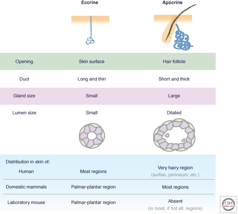
The human body is covered with several million sweat glands. These tiny coiled tubular skin appendages produce the sweat that is our primary source of cooling and hydration of the skin. Numerous studies have been published on their morphology and physiology. Until recently, however, little was known about how glandular skin maintains homeostasis and repairs itself after tissue injury. Here, we provide a brief overview of sweat gland biology, including newly identified reservoirs of stem cells in glandular skin and their activation in response to different types of injuries. Finally, we discuss how the genetics and biology of glandular skin has advanced our knowledge of human disorders associated with altered sweat gland activity.


Figures

Figure 1.
Figure 1.
Features of eccrine and apocrine sweat glands. (Image adapted from Sato et al. 1987.)
Figure 2.
Figure 2.
Development and morphology of mouse eccrine sweat glands. (A) Mouse sweat bud placodes start to form just before birth, and progenitors from the epidermal basal layer invaginate deep into dermis. As morphogenesis proceeds, cells at the tip of the down-growing sweat bud differentiate into luminal cells and myoepithelial cells of the secretory coil, whereas suprabasal cells in the duct extend upward and reach to the skin surface to form an orifice. (B) Cross-section and histology staining of a mouse paw pad where sweat apparatus are located, showing intraepidermal duct (IED), sweat duct (SD), and sweat gland (SG). (C) Ultrastructural image of a cross section of the sweat gland. Shown in purple are luminal cells containing clear cells and dark cells (asterisk). Myoepithelial cells are in green. Magnification of the boxed area is on the right, showing its spindle-like morphology, enrichment of actin filaments, and intimate contact with basement membrane. (Images courtesy of H.A. Pasolli.)
Figure 3.
Figure 3.
Homeostasis and wound repair of mouse eccrine sweat glands. (A, left) During homeostasis, basal cells in paw epidermis and sweat ducts proliferate to self-renew and replenish the worn suprabasal epidermis and intraepidermal duct. Curved arrows, self-renew; straight arrows, differentiation and migration. (A, middle) When a large portion of epidermis is injured or removed, increased proliferation takes place in neighboring healthy basal cells of the sweat duct and epidermis. These cells (and/or their progeny) rapidly migrate and differentiate to repair the injured area. Note that cells in the sweat gland do not respond to epidermal injury. (A, right) When cells in the sweat gland are injured, the neighboring healthy cells can be activated to repair locally, shown by curved arrows. (B) During glandular injury, when luminal cells are injured, neighboring luminal cells proliferate to repair (purple curved arrow). When myoepithelial cells are injured, neighboring myoepithelial cells proliferate to repair (green curved arrow). (C) Ultrastructural image of an unresponsive myoepithelial cell (green) in close vicinity of dying luminal cells (purple). Ap, apoptotic bodies. Scale bar, 1 µm (Lu et al. 2012).
Figure 4.
Figure 4.
De novo sweat gland formation by engraftment of myoepithelial cells purified from adult sweat glands. (A) K14H2BGFP myoepithelial cells purified from adult sweat glands (SG, left) and adult mammary glands (MG, right) show distinct morphology on engraftment. SG myoepithelial cells form a single luminal (K18+) layer and a small lumen; MG myoepithelial cells form multiple layers of luminal cells and an elongated lumen resembling the structure of terminal end buds. Yellow dashed lines outline the lumens. (B) Long-term graft of sweat gland myoepithelial cells form nonbranched, tightly tangled coil structure. Myoepithelial cells purified from Rosa26-YFP transgenic mice that allow lineage tracing gave rise to the entire structure, including K18+ luminal cells. (C) In a lactating host, grafts derived from sweat gland myoepithelial cells undergo transdifferentiation. (Left) Example of a graft that still expresses sweat gland marker (NKAα, ATP1a1) albeit lower than original level, but shows branching morphology. (Right) Example of a graft that loses NKAα expression, but starts to express milk protein (mammary gland marker) and its lumen is significantly enlarged. White solid line, basement membrane; yellow dashed line, lumen. Scale bars, 10 µm. (D) Ultrastructural analysis of a de novo glandular structure derived from purified sweat gland myoepithelial cells. Boxed areas are shown on the right. II, showing a myoepithelial cell, attaches to basement membrane (box a) and enriches in actin filament bundles (box b). III, magnification of a lumen, showing presence of microvilli (Mv). Intercellular junctions are boxed, showing desmosomes (De) and tight junction (Tj). Scale bars, 500 nm (Lu et al. 2012).

References

    1. Ashley I, Smith-Reed M, Chernys A 1997. Sweat gland carcinoma. Case report and review of the literature. Dermatol Surg 23: 129–133 - PubMed
    1. Biedermann T, Pontiggia L, Böttcher-Haberzeth S, Tharakan S, Braziulis E, Schiestl C, Meuli M, Reichmann E 2010. Human eccrine sweat gland cells can reconstitute a stratified epidermis. J Invest Dermatol 130: 1996–2009 - PubMed
    1. Biernat W, Peraud A, Wozniak L, Ohgaki H 1998. p53 mutations in sweat gland carcinomas. Int J Cancer 76: 317–320 - PubMed
    1. Blanpain C, Simons BD 2013. Unravelling stem cell dynamics by lineage tracing. Nat Rev Mol Cell Biol 14: 489–502 - PubMed
    1. Blanpain C, Lowry WE, Geoghegan A, Polak L, Fuchs E 2004. Self-renewal, multipotency, and the existence of two cell populations within an epithelial stem cell niche. Cell 118: 635–648 - PubMed

Publication types

MeSH terms