. 2014 Aug;60(2):576-87.
doi: 10.1002/hep.26996. Epub 2014 Jun 18.

Inactivation of Wnt signaling by a human antibody that recognizes the heparan sulfate chains of glypican-3 for liver cancer therapy

Affiliations

Inactivation of Wnt signaling by a human antibody that recognizes the heparan sulfate chains of glypican-3 for liver cancer therapy

Wei Gao et al. Hepatology. 2014 Aug.

Abstract

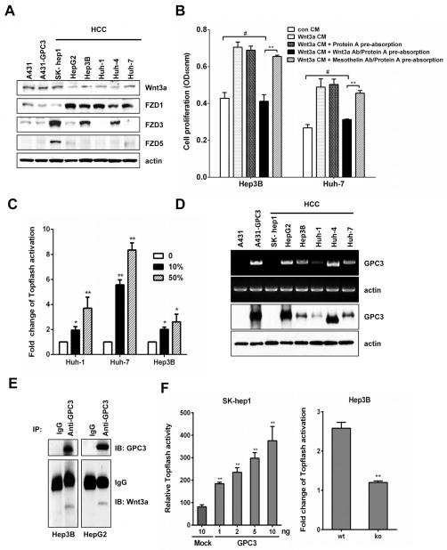
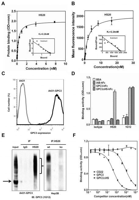
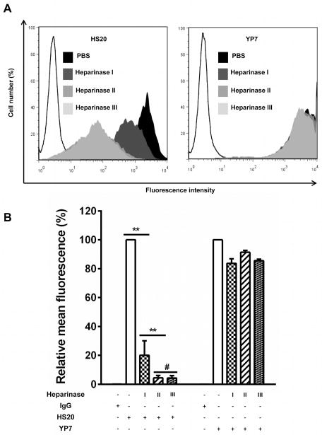
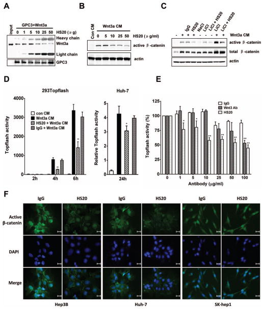
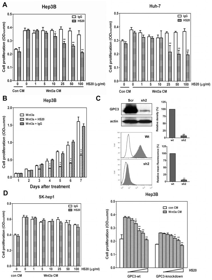
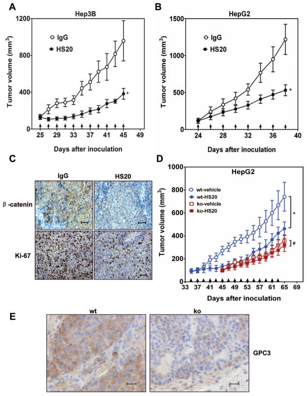
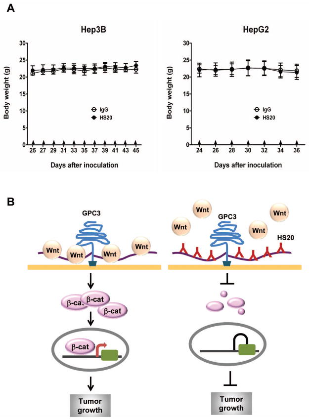
Wnt signaling is important for cancer pathogenesis and is often up-regulated in hepatocellular carcinoma (HCC). Heparan sulfate proteoglycans (HSPGs) function as coreceptors or modulators of Wnt activation. Glypican-3 (GPC3) is an HSPG that is highly expressed in HCC, where it can attract Wnt proteins to the cell surface and promote cell proliferation. Thus, GPC3 has emerged as a candidate therapeutic target in liver cancer. While monoclonal antibodies to GPC3 are currently being evaluated in preclinical and clinical studies, none have shown an effect on Wnt signaling. Here, we first document the expression of Wnt3a, multiple Wnt receptors, and GPC3 in several HCC cell lines, and demonstrate that GPC3 enhanced the activity of Wnt3a/β-catenin signaling in these cells. Then we report the identification of HS20, a human monoclonal antibody against GPC3, which preferentially recognized the heparan sulfate chains of GPC3, both the sulfated and nonsulfated portions. HS20 disrupted the interaction of Wnt3a and GPC3 and blocked Wnt3a/β-catenin signaling. Moreover, HS20 inhibited Wnt3a-dependent cell proliferation in vitro and HCC xenograft growth in nude mice. In addition, HS20 had no detectable undesired toxicity in mice. Taken together, our results show that a monoclonal antibody primarily targeting the heparin sulfate chains of GPC3 inhibited Wnt/β-catenin signaling in HCC cells and had potent antitumor activity in vivo.

Conclusion: An antibody directed against the heparan sulfate of a proteoglycan shows efficacy in blocking Wnt signaling and HCC growth, suggesting a novel strategy for liver cancer therapy.


Figures

Figure 1
Figure 1. Activation of Wnt signaling by GPC3 in HCC
(A) Wnt3a, FZD1, FZD3 and FZD5 expressions in HCC cell lines. (B) 50% Wnt3a CM was incubated with Wnt3a antibody or MSLN antibody and then the antibody-Wnt3a complex was removed by Protein A-Agarose beads. The pre-absorbed CM was added to Hep3B and Huh-7 cells. Cell proliferation was measured by WST-8 assay at 48 hours after treatment. CM: conditioned media, MSLN: mesothelin. The data represent the mean ± SD (**P <0.01 and #P > 0.05). (C) Topflash activity in Huh-1, Huh-7 and Hep3B cells. Twenty-four hours after transfection, cells were starved for 2 hours and then treated with 10% or 50% Wnt3a CM. Topflash activity was measured 24 hours later. The data represent the mean ± SD (*P <0.05 and **P <0.01). (D) GPC3 expression in HCC cell lines was measured by RT-PCR (top panel, showing full length GPC3) and Western blots (bottom panel, showing GPC3 core protein). (E) Immunoprecipitation assays detected the interaction of GPC3 and Wnt3a in Hep3B cells and HepG2 cells. The immunoprecipitated complex was detected with an anti-Wnt3a antibody or an anti-GPC3 antibody (YP7). (F) Topflash activity in GPC3-transfected SK-hep1 cells and GPC3 knockdown Hep3B cells. Twenty-four hours after transfection, cells were starved for 2 hours and then treated with 50% (SK-hep1 cells) or 100% Wnt3a CM (Hep3B cells). Topflash activity was measured 24 hours later in SK-hep1 cells and 48 hours in Hep3B cells; GPC3 knockdown efficiency was shown in Fig. 5C. Wt: wild type; ko: knockdown. The data represent the mean ± SD (**P <0.01).
Figure 2
Figure 2. Isolation of a human antibody targeting HS chains of GPC3
(A) The binding affinity of HS20 on GPC3-his. (B) The binding affinity of HS20 on A431-GPC3 cells. (C) Flow cytometry analysis of HS20 on A431 and A431-GPC3 cells. (D) Binding properties of HS20 on GPC3, GPC3ΔHS, or irrelevant human Fc protein IAB. 1G12, a commercial mouse antibody against GPC3. (E) Immunoprecipitation of GPC3 in A431-GPC3 cells, Hep3B, and Hep3B GPC3 knockdown cells. Cell lysates incubated with HS20 or human IgG for immunoprecipitation. The immunoprecipitated complex was detected by 1G12. Black arrow indicates the GPC3 core protein and the bracket indicates glycosylated GPC3. Wt: wild type; ko: knockdown. GPC3 knockdown efficiency has been shown in Fig. 5C. (F) Competitive ELISA. Different concentrations of GPC3 or GPC3ΔHS were pre-incubated with HS20 (0.1 nM) for 1 hour. The mixture was added into the GPC3-coated plate to measure the binding of HS20/GPC3. CD22 was used as the negative control. The data represent the mean ± SD.
Figure 3
Figure 3. Both sulfated and non-sulfated regions of HS chain are involved in HS20 binding
(A) Treatment of GPC3-expressing cells with heparinase I, II and III. A431-GPC3 cells were treated with different heparinases for 2 hours at 37°C. Cells were then stained with HS20 (5 μg/ml) or YP7 (1 μg/ml) for flow cytometry. (B) Statistical analysis of three independent experiments. The binding fluorescence signals of untreated cells represent 100%. The data represent the mean ± SD of three independent experiments (**P <0.01 and #P > 0.05).
Figure 4
Figure 4. HS20 blocks Wnt/β-catenin activation
(A) GPC3-hFc was pre-incubated with different amounts of HS20 for 1 hour on ice and then recombinant Wnt3a was added and incubated for another 2 hours on ice. Protein A-Agarose beads were added to capture the hFc fusion protein. The captured proteins were released with 1x SDS loading buffer. Western blot was performed to detect Wnt3a. (B) HEK293 SuperTopflash cells were starved for 2 hours and then pretreated with 2x indicated concentrations of HS20. An hour later, equal volumes of Wnt3a CM were added. Active β-catenin expression level was detected by Western blot 6 hours later. (C) HEK293 SuperTopFlash cells were starved for 2 hours and then pretreated with or without 200 μg/ml of HS20. One hour later, equal volumes of Wnt3a CM (combined with or without 20 mM LiCl or NaCl) were added. β-catenin expression level was detected 6 hours later. (D) HEK293 SuperTopflash cells or transfected Huh-7 cells were treated with 100 μg/ml or 25 ug/ml of HS20 as described in (B). Topflash activity was measured at indicated time points. The data represent the mean ± SD (*P <0.05 and **P <0.01). (E) HEK293 SuperTopflash cells were treated with different concentrations of human IgG, HS20 or anti-Wnt3a antibody as described in (B). Topflash activity was measured after 6 hours. The data represent the mean ± SD (*P <0.05 and **P <0.01). (F) Immunocytofluorescence staining of β-catenin in Hep3B, Huh-7 and SK-hep1 cells. Cells were treated as described in (B). Twenty-four hours later, cells were stained with active β-catenin antibody after fixation. Scale bar, 20 μm.
Figure 5
Figure 5. HS20 inhibits Wnt3a-induced cell proliferation
(A) Cells were starved for 2 hours and then pretreated with 2x indicated concentrations of HS20. One hour later, equal volumes of Wnt3a CM were added. Cells were cultured for 3 days and cell proliferation was measured by WST-8 assay. The data represent the mean ± SD (*p <0.05 and **P <0.01). (B) Hep3B cells were treated with 100 μg/ml of HS20 as described in (A). Cell proliferation was measured on different days by WST-8 assay. The data represent the mean ± SD (**P <0.01). (C) Wild type Hep3B cells and GPC3 knockdown Hep3B cells were treated with increased concentrations of HS20 as described in (A). Cell proliferation was measured by WST-8 assay after 3 days. GPC3 knockdown efficiency was calculated by western blot and flow cytometry. The data represent the mean ± SD (*P <0.05 and **P <0.01). (D) SK-hep1 cells were treated with 100 μg/ml of HS20 as described in (A). Cell proliferation was measured on day 3. The data represent the mean ± SD.
Figure 6
Figure 6. Anti-tumor activity via the HS20 antibody
BALB/c nu/nu mice were subcutaneously inoculated with 5×10⁶ Hep3B or HepG2 cells (A and B) or 3 × 10⁶ wild type or GPC3 knockdown HepG2 cells (C). When tumors reached an average volume of 100 mm3, mice were intravenously administered 20 mg/kg HS20 three times a week. The arrows indicate HS20 injections. The data represent the mean ± SD (*P <0.05 and #P >0.05). (A) Hep3B xenografts were treated with HS20 (n=5/group). Human IgG was injected as control. (B) HepG2 xenografts were treated with HS20 (n=7/group). Human IgG was injected as control. (C) β-catenin and Ki-67 immunohistochemistry staining of Hep3B xenografts. Scale bar, 100 μm. (D) Wild type HepG2 xenografts (n=5/group) and GPC3 knockdown HepG2 xenografts (n=4/group) were treated with HS20. PBS was used as vehicle control. (E) GPC3 expression level in wild type and knockdown xenograft were detected by YP7 staining.
Figure 7
Figure 7. Body weight of HS20-treated mice
(A) Body weight of HS20 treated mice. The data represent the mean ± SD (B) The working model for the mechanistic interaction of HS20 and GPC3. The numbers and locations of Wnt and antibody molecules bound to HS chains of GPC3 were drawn arbitrarily.

Comment in

References

    1. Ho M, Kim H. Glypican-3: a new target for cancer immunotherapy. Eur J Cancer. 2011;47:333–338. - PMC - PubMed
    1. Bengochea A, de Souza MM, Lefrancois L, Le Roux E, Galy O, Chemin I, Kim M, et al. Common dysregulation of Wnt/Frizzled receptor elements in human hepatocellular carcinoma. Br J Cancer. 2008;99:143–150. - PMC - PubMed
    1. Wei W, Chua MS, Grepper S, So SK. Blockade of Wnt-1 signaling leads to anti-tumor effects in hepatocellular carcinoma cells. Mol Cancer. 2009;8:76. - PMC - PubMed
    1. You XJ, Bryant PJ, Jurnak F, Holcombe RF. Expression of Wnt pathway components frizzled and disheveled in colon cancer arising in patients with inflammatory bowel disease. Oncol Rep. 2007;18:691–694. - PubMed
    1. Gurney A, Axelrod F, Bond CJ, Cain J, Chartier C, Donigan L, Fischer M, et al. Wnt pathway inhibition via the targeting of Frizzled receptors results in decreased growth and tumorigenicity of human tumors. Proc Natl Acad Sci U S A. 2012;109:11717–11722. - PMC - PubMed

Publication types

MeSH terms