Review
. 2014 Feb 3;369(1638):20130096.
doi: 10.1098/rstb.2013.0096. Print 2014 Mar 19.

Role of anoctamins in cancer and apoptosis

Affiliations
Review

Role of anoctamins in cancer and apoptosis

Podchanart Wanitchakool et al. Philos Trans R Soc Lond B Biol Sci. .

Abstract

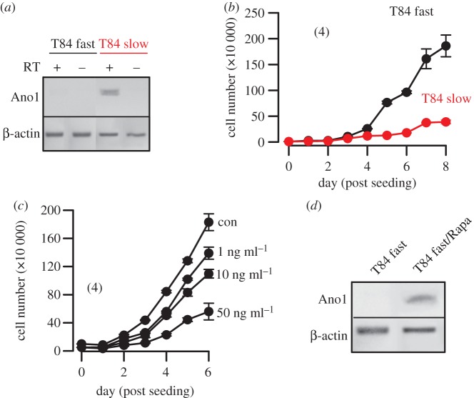
Anoctamin 1 (TMEM16A, Ano1) is a recently identified Ca(2+)-activated chloride channel and a member of a large protein family comprising 10 paralogues. Before Ano1 was identified as a chloride channel protein, it was known as the cancer marker DOG1. DOG1/Ano1 is expressed in gastrointestinal stromal tumours (GIST) and particularly in head and neck squamous cell carcinoma, at very high levels never detected in other tissues. It is now emerging that Ano1 is part of the 11q13 locus, amplified in several types of tumour, where it is thought to augment cell proliferation, cell migration and metastasis. Notably, Ano1 is upregulated through histone deacetylase (HDAC), corresponding to the known role of HDAC in HNSCC. As Ano1 does not enhance proliferation in every cell type, its function is perhaps modulated by cell-specific factors, or by the abundance of other anoctamins. Thus Ano6, by regulating Ca(2+)-induced membrane phospholipid scrambling and annexin V binding, supports cellular apoptosis rather than proliferation. Current findings implicate other cellular functions of anoctamins, apart from their role as Ca(2+)-activated Cl(-) channels.

Keywords: TMEM16A; TMEM16F; anoctamin 1; anoctamin 6; cancer; head and neck stromal cell carcinoma.


Figures

Figure 1.
Figure 1.
Ano1 controls proliferation: (a) Cell proliferation measured online by impedance-based xCELLigence proliferation assay system in HNSCC (BHY, CAL33) and colonic epithelial (HT29) cells (see electronic supplementary material). siRNA-knockdown of Ano1 expression reduced proliferation in both BHY and CAL33 cells, but slightly augmented proliferation in HT29 cells. Staurosporine (1 µg ml−1) strongly inhibited proliferation. (b) Attachment of BHY, CAL33 and H29 cells after seeding in xCELLigence chambers (see electronic supplementary material). (c) Migration of BHY, CAL33 and H29 cells after seeding in the xCELLigence migration chamber (see electronic supplementary material). (d) Summary of the rate of cell migration between 10 and 30 h after seeding and inhibition of migration by TA(10 µM). (e) Migration of CAL33 cells and effect of fetal calf serum and TA. Mean ± s.e.m. (number of cells). Symbol # denotes significant difference when compared with BHY and serum free, respectively (p < 0.05; ANOVA). Asterisks (*) denote significant inhibition by TA (p < 0.05; paired t-test). (Online version in colour.)
Figure 2.
Figure 2.
Anoctamins facilitate cell volume decrease and support diapedesis and cell migration: model for diapedesis and migration of a tumour cell expressing anoctamin Cl channels, which allow Cl release and osmotic cell shrinkage. Ano1 is regulated by actin and possibly by a number of proteins related to signalling/scaffolding, such as ezrin, radixin, moesin and proteins related to cell attachment and migration, such as zyxin, fibulin 1, S100A11, twinfilin and catenin. (Online version in colour.)
Figure 3.
Figure 3.
HDAC regulates expression of Ano1: (a,b) western blots and densitometric analysis indicating expression of Ano1 in HT29 colonic epithelial and BHY HNSCC cells, and inhibition by HDAC inhibitors valproic acid (3 mM) and butyrate (4 mM). ß-actin was used as a loading control. (c) Real-time PCR analysis of Ano1-mRNA expression in HT29 and BHY cells. (d,e) Induction of apoptosis and inhibition of proliferation of BHY cells by valproic acid and butyric acid, as measured by apoptosis assays (see electronic supplementary material) and cell counting. (f) Whole cell Ano1 Cl currents (Vc = +100 mV) activated by an increase in intracellular Ca2+ owing to stimulation with the purinergic agonist ATP (100 µM). (g) Expression of Ano1 in human HNSCC samples and normal tissue as measured by immunohistochemistry (cf. electronic supplementary material). (h) Inhibition of proliferation of UM-SCC cancer cells by various concentrations of TSA. Mean ± s.e.m. (number of cells). Symbol # denotes significant difference when compared with control, normal tissue, HT29 cells or absence of HDAC inhibitors, respectively (p < 0.05; unpaired t-test).
Figure 4.
Figure 4.
APC/mTOR controls expression of Ano1 in mouse intestine: (a,b) western blot analysis of expression of Ano1 in duodenum, ileum and colon of APCmin/+ and wild-type mice. (c) Semiquantitiative analysis (see electronic supplementary material) of Ano1-mRNA expression in duodenum, ileum and colon of APCmin/+ and wild-type mice. (d) Treatment with the mTOR-inhibitor rapamycin [51] increased expression of Ano1 in the intestine of APCmin/+ mice. Mice were fed 40 mg kg−1 Sirolimus (Wyeth Pharmaceuticals, Collegeville, PA, USA) in their chow to reach blood levels of 12 ± 2 ng ml−1 rapamycin (n = 66). Mean ± s.e.m. (number of cells). Symbol # denotes significant difference when compared with +/+ or –Rapa, respectively (p < 0.05; unpaired t-test).
Figure 5.
Figure 5.
APC/mTOR controls expression of Ano1 and proliferation of T84 cells: (a) RT-PCR analysis of Ano1-mRNA expression in fast growing and slowly growing T84 colonic carcinoma cells. +/− RT indicates the presence of or the absence of reverse transcriptase in the reaction. (b) Growth curves for fast growing and slowly growing T84 cells. (c) Effect of treatment with rapamycin on proliferation of T84 fast cells. (d) RT-PCR analysis of Ano1-mRNA expression in fast growing T84 cells in the absence and the presence of rapamycin. The rapamycin concentration used was 50 ng ml−1. Mean ± s.e.m. (number of cells). (Online version in colour.)

Similar articles

Cited by

References

    1. AlDehni F, Spitzner M, Martins JR, Barro Soria R, Schreiber R, Kunzelmann K. 2009. Role of bestrophin for proliferation and in epithelial to mesenchymal transition. J. Am. Soc. Nephrol. 20, 1556–1564. (10.1681/ASN.2008090987) - DOI - PMC - PubMed
    1. Caputo A, et al. 2008. TMEM16A, a membrane protein associated with calcium-dependent chloride channel activity. Science 322, 590–594. (10.1126/science.1163518) - DOI - PubMed
    1. Ousingsawat J, Martins JR, Schreiber R, Rock JR, Harfe BD, Kunzelmann K. 2009. Loss of TMEM16A causes a defect in epithelial Ca2+ dependent chloride transport. J. Biol. Chem. 284, 28 698–28 703. (10.1074/jbc.M109.012120) - DOI - PMC - PubMed
    1. Schroeder BC, Cheng T, Jan YN, Jan LY. 2008. Expression cloning of TMEM16A as a calcium-activated chloride channel subunit. Cell 134, 1019–1029. (10.1016/j.cell.2008.09.003) - DOI - PMC - PubMed
    1. Almaca J, Tian Y, AlDehni F, Ousingsawat J, Kongsuphol P, Rock JR, Harfe BD, Schreiber R, Kunzelmann K. 2009. TMEM16 proteins produce volume regulated chloride currents that are reduced in mice lacking TMEM16A. J. Biol. Chem. 284, 28 571–28 578. (10.1074/jbc.M109.010074) - DOI - PMC - PubMed

Publication types

MeSH terms