Review
. 2014 Feb 3;369(1638):20130105.
doi: 10.1098/rstb.2013.0105. Print 2014 Mar 19.

Regulation of voltage-gated sodium channel expression in cancer: hormones, growth factors and auto-regulation

Affiliations
Review

Regulation of voltage-gated sodium channel expression in cancer: hormones, growth factors and auto-regulation

Scott P Fraser et al. Philos Trans R Soc Lond B Biol Sci. .

Abstract

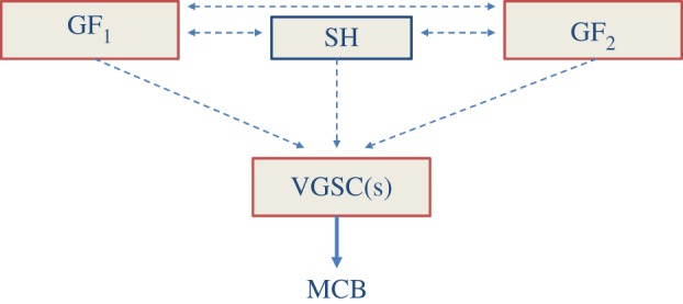
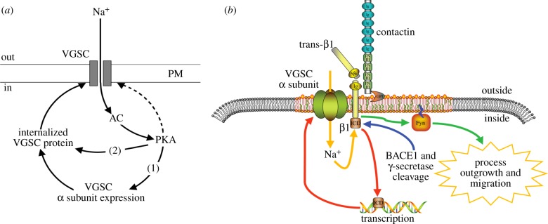
Although ion channels are increasingly being discovered in cancer cells in vitro and in vivo, and shown to contribute to different aspects and stages of the cancer process, much less is known about the mechanisms controlling their expression. Here, we focus on voltage-gated Na(+) channels (VGSCs) which are upregulated in many types of carcinomas where their activity potentiates cell behaviours integral to the metastatic cascade. Regulation of VGSCs occurs at a hierarchy of levels from transcription to post-translation. Importantly, mainstream cancer mechanisms, especially hormones and growth factors, play a significant role in the regulation. On the whole, in major hormone-sensitive cancers, such as breast and prostate cancer, there is a negative association between genomic steroid hormone sensitivity and functional VGSC expression. Activity-dependent regulation by positive feedback has been demonstrated in strongly metastatic cells whereby the VGSC is self-sustaining, with its activity promoting further functional channel expression. Such auto-regulation is unlike normal cells in which activity-dependent regulation occurs mostly via negative feedback. Throughout, we highlight the possible clinical implications of functional VGSC expression and regulation in cancer.

Keywords: activity-dependent regulation; growth factor; hormone; metastasis; voltage-gated sodium channel.


Figures

Figure 1.
Figure 1.
Schematic diagram of the structure and membrane topology of the voltage-gated sodium channel showing the main regulatory sites. Given α-subunits have four domains (DI–DIV) each composed of six transmembrane segments. Within the latter, segment four contains positively charged amino acids and this is the main voltage-sensitive region; the loop between transmembrane segments 5 and 6 is negatively charged and forms the pore region. Many types of modulatory sites exist for both α- and β-subunits as indicated by the key. The boxes adjacent to α- and β-subunits list the proteins known to interact with each subunit, respectively. PDZ, post-synaptic density protein (PSD95) and Drosophila disc large tumour suppressor (Dlg1) and zonula occludens-1 protein (zo-1); ER, endoplasmic reticulum; RXR, a motif which mediates retention of proteins in the ER. (Online version in colour.)
Figure 2.
Figure 2.
Summary diagrams showing possible regulatory pathways controlling voltage-gated sodium channel (VGSC) expression/activity in cells by (a) hormones and (b) growth factors. Some pathways mediate transcriptional activity; some actions are through the respective receptors, whereas some actions are directly on the channel. Some of the regulatory pathways have only been described in normal cells. Abbreviations are defined in text. VGSC↓ and VGSC↑ denote decreased and increased VGSC expression/activity, respectively. (Online version in colour.)
Figure 3.
Figure 3.
Effects of oestrogen receptor α (ERα) on voltage-gated sodium channel (VGSC) expression and activity in MDA-MB-231 cells transfected with ERα (MDA-MB-231-ERα cells). (a) Basal levels of nNav1.5 mRNA were significantly lower in MDA-MB-231-ERα cells (ERα) compared with control cells expressing only the plasmid vector (VC5; p < 0.001; n = 4). (b) Treatment of MDA-MB-231-ERα cells with the ER antagonist ICI-182,780 (ICI; 1 µM) for more than 72 h significantly increased the nNav1.5 mRNA level, compared with non-treated cells (Cntl; p < 0.001; n = 4). (c) Similar treatment with ICI-182 780 for more than 48 h significantly increased the number of cells with VGSC activity, in comparison with those grown in normal medium (5% FBS–Cntl) as determined by patch-clamp recording (Fisher's exact test: p < 0.001; n = 73 cells). (d) Treatment of MDA-MB-231-ERα cells with ICI-182,780 for more than 72 h significantly increased the lateral motility of the cells (p < 0.01; n = 4). Further information is given in the supplementary Methods section.
Figure 4.
Figure 4.
Upregulation of functional expression of Nav1.7 in human non-small-cell lung cancer H460 cells and consequent increase in invasiveness via ERK1/2 signalling. (a) Current–voltage (I–V) plots for control/untreated cells (open squares) and cells treated in the presence of serum for 24 h with 100 ng ml−1 EGF (closed squares), 1 μM gefitinib/Gef (open circles) or 10 μg ml−1 EGF receptor blocking antibody (filled circles). Currents were evoked using 30 ms depolarizing steps in 5 mV intervals (−90 to +70 mV) from a holding potential of −90 mV. (b) I–V plots for control/untreated cells (open squares) and cells treated with 10 μM U0126 (closed squares). (c) Relative Nav1.7 mRNA expression showing effect of serum starvation for 48 h and treatment for 24 h with EGF (100 ng ml−1), Gef (1 μM) and co-application of EGF + Gef. (d) Matrigel invasion measured after 48 h in control medium (CTL), 0.5 μM TTX, 100 ng ml−1 EGF, 1 μM Gef and EGF + TTX. (e) Effects of treatment with 10 μM U0126 or 100 nM wortmannin (WORT) for 24 h on relative Nav1.7 mRNA expression, compared with control/untreated (CTL) cells. (f) Matrigel invasion measured over 48 h in control/untreated cells (CTL), and following treatment with 10 μM U0126, 10 μM U0126/1 μM TTX, 100 nM WORT, and WORT + TTX. (g) Proposed model for EGF-mediated upregulation of Nav1.7 and consequent invasiveness of H460 cells. Stimulation of EGFR with EGF results in increased functional expression of Nav1.7 via ERK1/2. Following transcription and translation, the mature Nav1.7 protein is trafficked to the cell surface where it becomes functional. At the resting membrane potential, VGSCs are partially activated but not fully inactivated, resulting in a basal influx of Na+. This increase in [Na+]i then drives cell invasion through an, as yet, unknown mechanism. All data are presented as means ± s.e. (n = 6–13). Statistical analyses were with Student's t-test or one-way ANOVA and Student–Newman–Keuls correction, as appropriate; significance: *p < 0.05, **p < 0.01, ***p < 0.001. Adapted from [89]. (Online version in colour.)
Figure 5.
Figure 5.
A ‘conceptual’ scheme showing how growth factors (GF1, GF2, etc.) and steroid hormone (SH) signalling systems can feed through and compensate for each other in regulating expression/activity of the VGSC(s). In turn, VGSC activity enhances metastatic cell behaviour (MCB). Dotted, horizontal lines denote the interactive pathways, involving mostly the intracellular signalling cascades. (Online version in colour.)
Figure 6.
Figure 6.
Activity-dependent regulation of VGSC expression and VGSC-dependent migration. (a) In Mat-LyLu [28] and MDA-MB-231 [34] cells, INa activates adenylate cyclase (AC) and protein kinase A (PKA), which in turn (i) potentiates α-subunit mRNA expression and (ii) increases channel expression at the plasma membrane, without affecting total cellular α-subunit protein level. PKA also directly phosphorylates surface-expressed VGSCs, although this may be independent of PKA (dashed line). Adapted from [28]. (b) Interplay between α and β1 subunits in transcription and process outgrowth, modelled from cerebellar granule neurons. Trans adhesion between β1 on an adjacent cell and a VGSC signalling complex (comprising α, β1 subunits and contactin), initiates a signalling cascade via FYN kinase that enhances process outgrowth and migration. Proteolytic processing of β1 by BACE1 and γ-secretase is proposed to release the soluble intracellular domain of β1, which may in turn enhance transcription of α subunit genes. Nav1.6 activity is required for β1-mediated process outgrowth, and in turn, β1 is required for normal localization of α-subunits. Thus, INa may fine-tune the dual processes of gene expression and migration. Adapted from [117]. (Online version in colour.)

Similar articles

Cited by

References

    1. Brackenbury WJ. 2012. Voltage-gated sodium channels and metastatic disease. Channels 6, 352–361. (10.4161/chan.21910) - DOI - PMC - PubMed
    1. Djamgoz MBA. 2011. Bioelectricity of cancer: voltage-gated ion channels and direct-current electric fields. In The physiology of bioelectricity in development, tissue regeneration, and cancer (ed. Pullar C.), pp. 269–294. London, UK: Taylor & Francis.
    1. Fraser SP, Pardo LA. 2008. Ion channels: functional expression and therapeutic potential in cancer. Colloquium on ion channels and cancer. EMBO Rep. 9, 512–515. (10.1038/embor.2008.75) - DOI - PMC - PubMed
    1. Pedersen SF, Stock C. 2013. Ion channels and transporters in cancer: pathophysiology, regulation and clinical potential. Cancer Res. 73, 1658–1661. (10.1158/0008-5472.CAN-12-4188) - DOI - PubMed
    1. Djamgoz MBA, Onkal R. 2013. Persistent current blockers of voltage-gated sodium channels: a clinical opportunity for controlling metastatic disease. Recent Patents Anti-Cancer Drug Discov. 8, 66–84. - PubMed

Publication types

MeSH terms

Substances