. 2013 Jan 1;5(1):222-34.
doi: 10.4161/derm.24808.

A pilot study assessing the effect of prolonged administration of high daily doses of vitamin D on the clinical course of vitiligo and psoriasis

Affiliations

A pilot study assessing the effect of prolonged administration of high daily doses of vitamin D on the clinical course of vitiligo and psoriasis

Danilo C Finamor et al. Dermatoendocrinol. .

Abstract

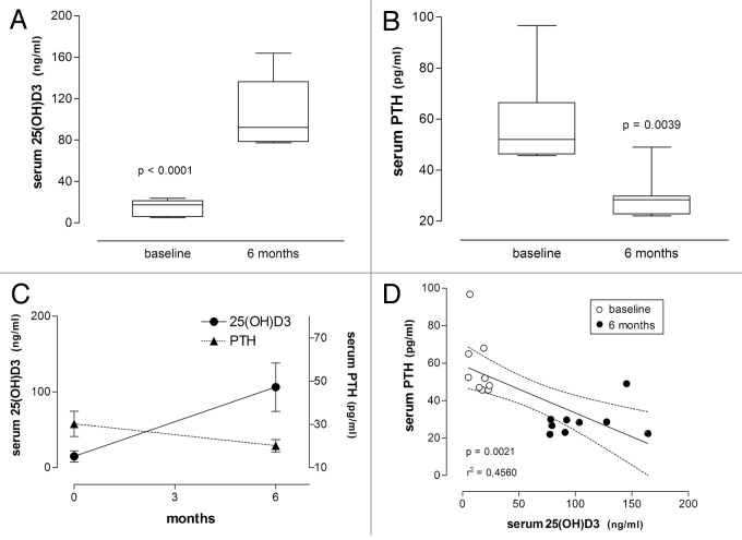
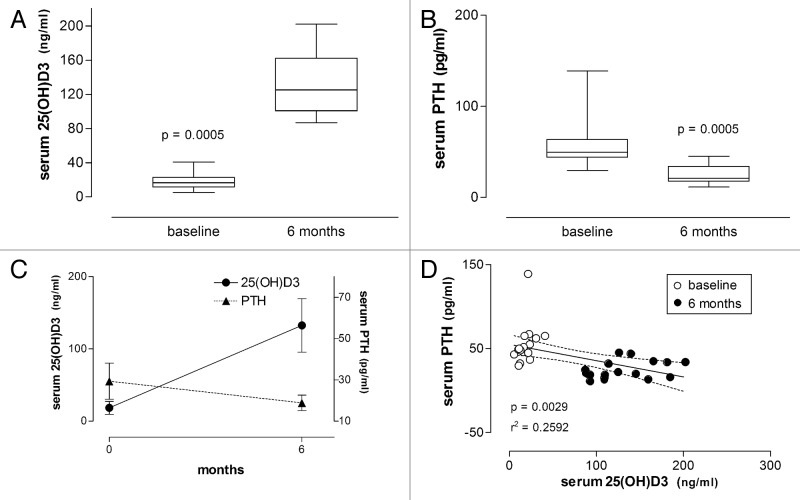
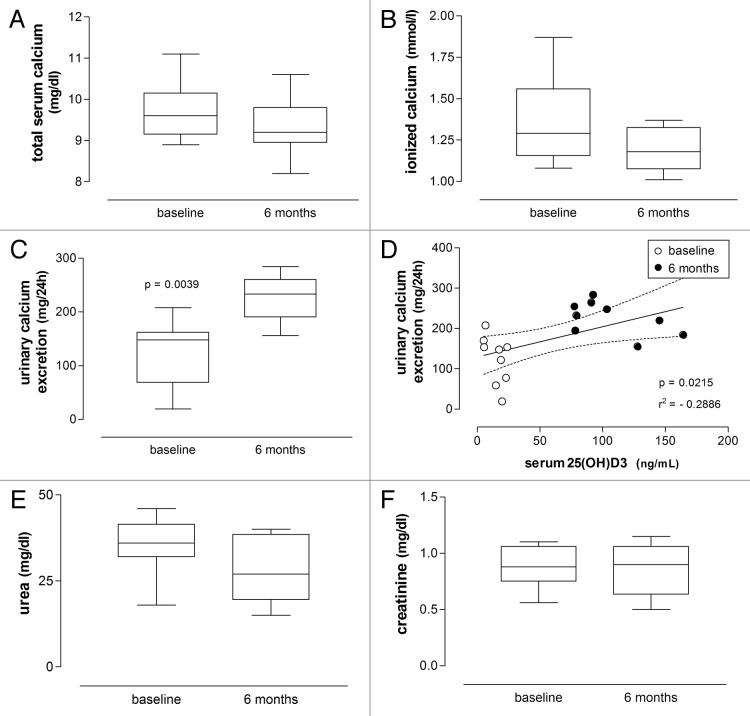
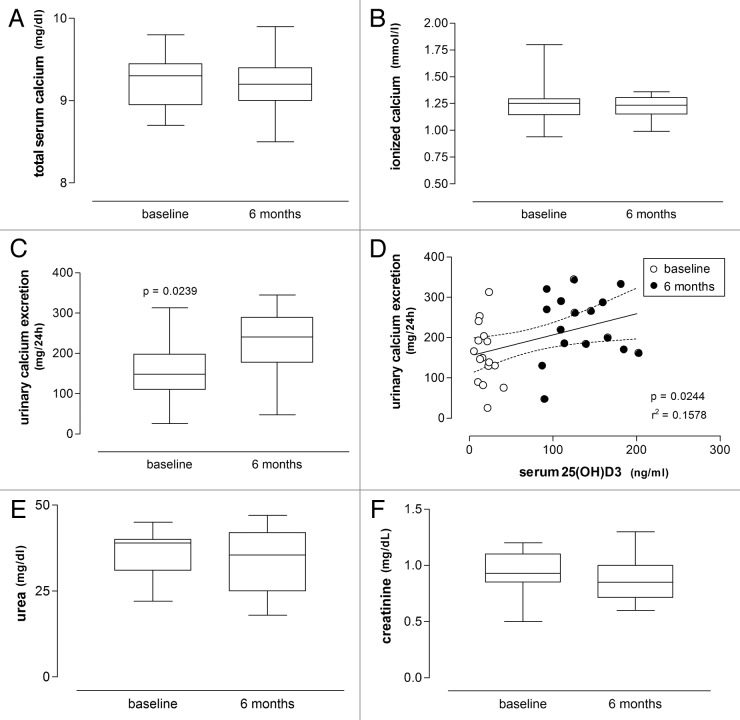
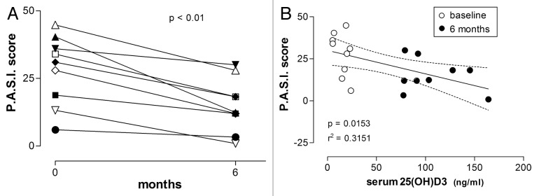
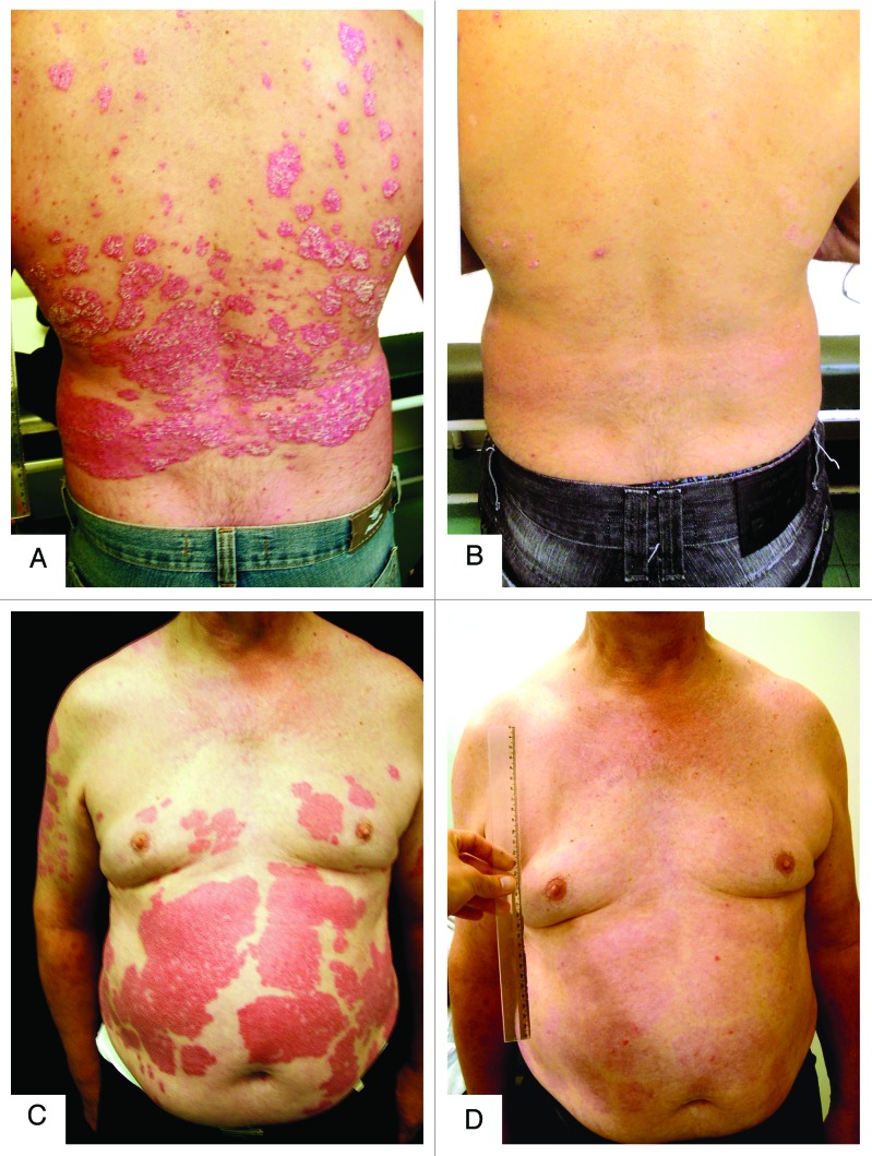
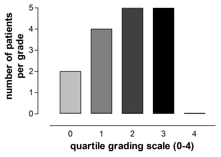
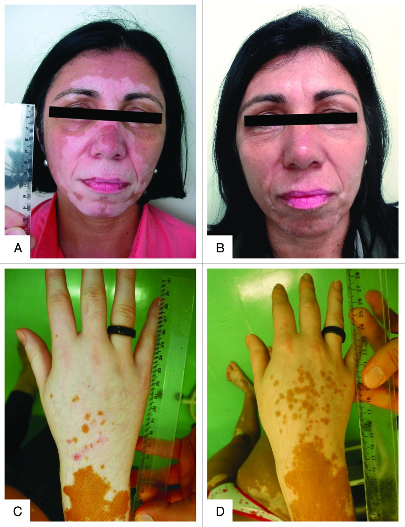
Autoimmunity has been associated with vitamin D deficiency and resistance, with gene polymorphisms related to vitamin D metabolism frequently described in affected patients. High doses of vitamin D3 may conceivably compensate for inherited resistance to its biological effects. This study aimed to assess the efficacy and safety of prolonged high-dose vitamin D3 treatment of patients with psoriasis and vitiligo. Nine patients with psoriasis and 16 patients with vitiligo received vitamin D3 35,000 IU once daily for six months in association with a low-calcium diet (avoiding dairy products and calcium-enriched foods like oat, rice or soya "milk") and hydration (minimum 2.5 L daily). All psoriasis patients were scored according to "Psoriasis Area and Severity Index" (PASI) at baseline and after treatment. Evaluation of clinical response of vitiligo patients required a quartile grading scale. All patients presented low vitamin D status (serum 25(OH)D3 ≤ 30 ng/mL) at baseline. After treatment 25(OH)D3 levels significantly increased (from 14.9 ± 7.4 to 106.3 ± 31.9 ng/mL and from 18.4 ± 8.9 to 132.5 ± 37.0 ng/mL) and PTH levels significantly decreased (from 57.8 ± 16.7 to 28.9 ± 8.2 pg/mL and from 55.3 ± 25.0 to 25.4 ± 10.7 pg/mL) in patients with psoriasis and vitiligo respectively. PTH and 25(OH)D3 serum concentrations correlated inversely. The PASI score significantly improved in all nine patients with psoriasis. Fourteen of 16 patients with vitiligo had 25-75% repigmentation. Serum urea, creatinine and calcium (total and ionized) did not change and urinary calcium excretion increased within the normal range. High-dose vitamin D3 therapy may be effective and safe for vitiligo and psoriasis patients.

Keywords: 25(OH)D3; autoimmunity; calcium; high dose; psoriasis; toxicity; treatment; vitamin D; vitiligo.


Figures

None
Figure 1. Enzyme kinetics of three hypothetical polymorphic variants of 25-hydroxyvitamin D-1 α-hydroxylase (CYP27B1), “A,” “B” and “C.” Some variants predictably exhibit decreased affinity for the substrate (increased Km) and/or decreased Vmax, requiring requiring a supra-physiologic substrate concentration to achieve a physiologic rate of substrate transformation. The polymorphic variants “B” and “C” require higher plasma concentrations of 25-hydroxyvitamin D (“x2” and “x3” respectively) to achieve the rate “y” of 1,25-dihydroxy vitamin D synthesis than the Km (“x1”) of the enzyme variant “A.” Compared with “A” or “B,” the polymorphic variant “C” requires a much higher plasma concentration of 25-hydroxyvitamin D to compensate for a reduced intracellular activity and achieve physiologic concentrations of 1,25-dihydroxy vitamin D within the immune cells.
None
Figure 2. Serum concentrations of 25(OH)D3 and PTH in patients with psoriasis before and after treatment with vitamin D (35,000 IU per day for 6 mo). (A) Box plot showing serum concentrations of 25(OH)D3 before and after treatment. (B) Same for the respective serum PTH concentrations. Significance level (Wilcoxon signed rank test) indicated in (A) and (B). (C) Serum concentrations of 25(OH)D3 and PTH respectively increased and decreased during treatment. (D) Linear regression of serum PTH on serum 25(OH)D3 concentrations is significant (significance level and r2 value are shown; dashed lines represent the 95% CIs for the linear regression line; baseline and 6-mo values are respectively shown as empty and filled circles).
None
Figure 5. Serum concentrations of 25(OH)D3 and PTH in patients with vitiligo before and after treatment with vitamin D (35,000 IU per day for 6 mo). (A) Box plot showing serum concentrations of 25(OH)D3 before and after treatment. (B) Same for the respective serum PTH concentrations. Significance level (Wilcoxon signed rank test) indicated in (A) and (B). (C) Serum concentrations of 25(OH)D3 and PTH respectively increased and decreased during treatment. (D) Linear regression of serum PTH on serum 25(OH)D3 concentrations is significant (significance level and r2 value are shown; dashed lines represent the 95% CIs for the linear regression line; baseline and 6-mo values are respectively shown as empty and filled circles).
None
Figure 3.(A), (B), (C), (D), (F) Box plots respectively showing concentrations of serum calcium, 24-h urinary calcium excretion, serum urea and serum creatinine in patients with psoriasis before and after treatment with vitamin D (35,000 IU per day for 6 mo), with a significant pre- and post-treatment difference only found for 24-h urinary calcium excretion (Wilcoxon signed rank test). (D) Linear regression of 24-h urinary calcium excretion on serum 25(OH)D3 concentrations is significant (significance level and r2 value are shown; dashed lines represent the 95% CIs for the linear regression line; baseline and 6-mo values are respectively shown as empty and filled circles).
None
Figure 6.(A), (B), (C), (D), (F) Box plots respectively showing concentrations of serum calcium, 24-h urinary calcium excretion, serum urea and serum creatinine in patients with vitiligo before and after treatment with vitamin D (35,000 IU per day for 6 mo), with a significant pre- and post-treatment difference only found for 24-h urinary calcium excretion (Wilcoxon signed rank test). (D) Linear regression of 24-h urinary calcium excretion on serum 25(OH)D3 concentrations is significant (significance level and r2 value are shown; dashed lines represent the 95% CIs for the linear regression line; baseline and 6-mo values are respectively shown as empty and filled circles).
None
Figure 4. PASI scores in patients with psoriasis before and after treatment with vitamin D (35,000 IU per day for 6 mo). (A) Individual temporal profiles of the P.A.S.I. score during the treatment showing clinical improvement in all patients. (B) Linear regression of P.A.S.I. on 25(OH)D3 concentration is significant (significance level and r2 value are shown; dashed lines represent the 95% CIs for the linear regression line; baseline and 6-mo values are respectively shown as empty and filled circles).
None
Figure 8. Photographs of two male patients with psoriasis before (A and C) and after (B and D) six months of treatment with vitamin D (35,000 IU per day). (A and B) A 59 y-old patient with BMI of 24.8 presenting a PASI score of 31 before treatment and achieving score of 18.2 after six months of treatment; his serum concentration of 25(OH)D3 was 22.8 ng/mL at baseline, reaching 127.5 ng/mL after 6 mo of treatment. (C and D) 60 y-old patient with BMI of 33.6 presenting a PASI score of 40.4 at baseline, achieving score of 12.4 after six months; his serum concentration of 25(OH)D3 was 5.6 ng/mL, reaching 103.2 ng/mL after six months of treatment.
None
Figure 7. Clinical response of 16 patients with vitiligo after treatment with vitamin D (35,000 IU per day for six months). Two patients showed no repigmentation (quartile 0); four patients showed 1‒25% repigmentation (quartile 1), five patients showed 26–50% repigmentation (quartile 2), five patients showed 51‒75% repigmentation (quartile 3) and none showed more than 75% repigmentation (quartile 4).
None
Figure 9. Photographs of two female patients with vitiligo before (A and C) and after (B and D) six months of treatment with vitamin D (35,000 IU per day). (A and B) A 50 y-old patient with BMI of 28.3 achieving between 51 and 75% of repigmentation (quartile 3) after six months of treatment; her serum concentration of 25(OH)D3 was 12.5 ng/mL, at baseline, reaching 92.4 ng/mL after 6 mo of treatment. (C and D) A 36 y-old patient with BMI of 22.7 achieving between 1 and 25% of repigmentation (quartile 1) after six months of treatment; her serum concentration of 25(OH)D3 was 12.0 ng/mL, at baseline, reaching 92.5 ng/mL after 6 mo of treatment.

Similar articles

Cited by

References

    1. Ramagopalan SV, Heger A, Berlanga AJ, Maugeri NJ, Lincoln MR, Burrell A, et al. A ChIP-seq defined genome-wide map of vitamin D receptor binding: associations with disease and evolution. Genome Res. 2010;20:1352–60. doi: 10.1101/gr.107920.110. - DOI - PMC - PubMed
    1. Holick MF. The vitamin D deficiency pandemic and consequences for nonskeletal health: mechanisms of action. Mol Aspects Med. 2008;29:361–8. doi: 10.1016/j.mam.2008.08.008. - DOI - PMC - PubMed
    1. Eyles DW, Smith S, Kinobe R, Hewison M, McGrath JJ. Distribution of the vitamin D receptor and 1 alpha-hydroxylase in human brain. J Chem Neuroanat. 2005;29:21–30. doi: 10.1016/j.jchemneu.2004.08.006. - DOI - PubMed
    1. Adams JS, Hewison M. Extrarenal expression of the 25-hydroxyvitamin D-1-hydroxylase. Arch Biochem Biophys. 2012;523:95–102. doi: 10.1016/j.abb.2012.02.016. - DOI - PMC - PubMed
    1. Eyles DW, Burne TH, McGrath JJ. Vitamin D, effects on brain development, adult brain function and the links between low levels of vitamin D and neuropsychiatric disease. Front Neuroendocrinol. 2013;34:47–64. doi: 10.1016/j.yfrne.2012.07.001. - DOI - PubMed

LinkOut - more resources