Review
. 2014 Feb;39(3):508-19.
doi: 10.1111/ejn.12462.

The neurobiology of skeletal pain

Affiliations
Review

The neurobiology of skeletal pain

Patrick W Mantyh. Eur J Neurosci. 2014 Feb.

Abstract

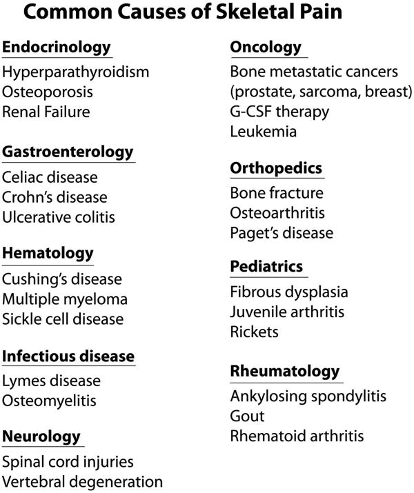
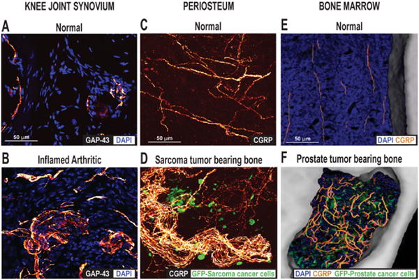
Disorders of the skeleton are one of the most common causes of chronic pain and long-term physical disability in the world. Chronic skeletal pain is caused by a remarkably diverse group of conditions including trauma-induced fracture, osteoarthritis, osteoporosis, low back pain, orthopedic procedures, celiac disease, sickle cell disease and bone cancer. While these disorders are diverse, what they share in common is that when chronic skeletal pain occurs in these disorders, there are currently few therapies that can fully control the pain without significant unwanted side effects. In this review we focus on recent advances in our knowledge concerning the unique population of primary afferent sensory nerve fibers that innervate the skeleton, the nociceptive and neuropathic mechanisms that are involved in driving skeletal pain, and the neurochemical and structural changes that can occur in sensory and sympathetic nerve fibers and the CNS in chronic skeletal pain. We also discuss therapies targeting nerve growth factor or sclerostin for treating skeletal pain. These therapies have provided unique insight into the factors that drive skeletal pain and the structural decline that occurs in the aging skeleton. We conclude by discussing how these advances have changed our understanding and potentially the therapeutic options for treating and/or preventing chronic pain in the injured, diseased and aged skeleton.

Keywords: CRPS; age; bone; cartilage; marrow; nociceptor.


Figures

Figure 1
Figure 1
List of human disorders that are frequently accompanied by skeletal pain. A major reason that skeletal pain occurs in such a diverse group of disorders is that the skeleton participates in a variety of functions including structural support, ambulation, protection of internal organs, mineral and growth factor storage and release, and the birth and maturation of blood cells. As the skeleton is composed of tissues with very unique microenvironments, (such as articular cartilage, periosteum, mineralized bone, and bone marrow) injury, aging or disease in any of these compartments can result in skeletal pain. For an extensive list of diseases that are frequently accompanied by skeletal pain, see http://www.rightdiagnosis.com/symptoms/bone_pain/common.htm in children, http://www.nof.org/articles/5 and for a list of rare (orphan) bone diseases that may be accompanied by skeletal pain, http://www.usbji.org/projects/RBDPN_op.cfm?dirID=252.
Figure 2
Figure 2
Changes in the skeleton and sclerostin levels with normal aging. (A) Human bone mass usually peaks at 25-30 years of age in both men and women and then generally declines thereafter, (B) resulting in stereotypic changes in posture and height. (C) One factor that appears to participate in driving age-related bone loss is sclerostin (here measured in plasma) which inhibits bone formation and increases linearly with age(D) The major cell type that expresses sclerostin in the adult is the osteocyte in bone, and sclerostin appears to exert its inhibitor effect by blocking bone progenitor cells and osteoblast function. (E) Administration of an antibody that sequesters sclerostin (Scl-Ab) has been shown to accelerate fracture healing in the primate tibia (reprinted with permission from Ominsky et al., 2011)(F) Administration of Scl-Ab to normal rats also shows a dose-related effect (as analyzed by μCT) in stimulating bone formation in the femoral neck of the femur, distal head of the femur and the L5 vertebrae (Figure reprinted with permission from Li et al., 2010).
Figure 3
Figure 3
The types of cells in bone, algogenic factors that can be released from these cells, and the primary afferent sensory nerve fibers that innervate the bone. (A) Schematic diagram illustrating the general organization and pattern of sensory innervation of the skeleton. Note that the relative density of A-delta and C sensory fibers (nerve fibers per unit area) is greatest in the periosteum, followed by the bone marrow and cortical bone, with a ratio of 100: 2: 0.1, respectively. (B) Primary afferent neurons innervating the skeleton have their cell bodies in the dorsal root ganglia (DRG) and project to the spinal cord. The types of sensory neurons that innervate the bone are unmyelinated C fibers and thinly myelinated Aδ fibers. The great majority (>80%) of sensory nerve fibers that innervate the bone and articular cartilage express TrkA (which is the cognate receptor for NGF), whereas < 30% of the nerve fibers that innervate the skin express TrkA. (C) Bone contains a remarkably diverse population of stromal, myeloid and other cells which can release a wide variety of factors (some of which are listed in the arrow) that can activate and sensitize the primary afferent nerve fibers that innervate the bone.
Figure 4
Figure 4
The efficacy of anti-NGF in blocking skeletal pain in pre-clinical and human models. (A) Radiograph of the normal mouse bone with intramedullary pin, and following a closed femoral fracture that generates fracture pain. (B) Sustained anti-NGF therapy (commenced immediately after fracture) attenuated fracture-induced skeletal pain-related guarding behaviors by 40-50%. Figure reprinted with permission from Koewler et al. (2007). (C) Radiograph of the normal and osteoarthritic human knee. (D) The assessment of the patient's knee pain while walking showed a significant reduction (40-50%) with human anti-NGF therapy (Tanezumab). A decrease in the change from baseline indicates an improvement (i.e., less pain). Figure reprinted with permission from Lane et al. (2010).
Figure 5
Figure 5
Ectopic sprouting of primary afferent sensory nerve fibers occurs in a variety of skeletal pain conditions. (A) Confocal images of sections of (A) the normal and (B) the inflamed knee joint that have been immunostained for DAPI, which stains nuclei, and growth-associated protein (GAP-43), which stains sprouting nerve fibers. Twenty-eight days after the initial injection of Complete Freund's Adjuvant (CFA) into the rat knee-joint, a significant number of GAP-43+ nerve fibers in the synovial knee joint had sprouted and had a disorganized appearance, as compared with vehicle-injected mice. Reprinted with permission from Jimenez-Andrade & Mantyh (2012). Confocal images of periosteal whole mounts of (C) normal and (D) sarcoma tumor-bearing bone immunostained for calcitonin gene-related peptide (CGRP+) and green fluorescent protein (GFP)-labeled sarcoma. As tumor cells invade the periosteum of the bone, ectopic sprouting of CGRP+ sensory fibers occurs and neuroma-like structures form. Reprinted with permission from Mantyh et al. (2010). Confocal images of bone marrow of (E) normal and (F) prostate tumor-bearing bone marrow, immunostained for DAPI, CGRP and GFP-expressing prostate cancer cells. Note that in the normal mice, CGRP+ nerve fibers present in the marrow space of normal mice appear as single nerve fibers with a highly linear morphology. As GFP+ prostate tumor cells proliferate and form tumor colonies, the CGRP+ sensory nerve fibers undergo marked sprouting which produces a highly branched, disorganized and dense meshwork of sensory nerve fibers that is never observed in normal marrow. Reprinted with permission from Jimenez-Andrade et al. (2010).

References

    1. Albrecht PJ, Hines S, Eisenberg E, Pud D, Finlay DR, Connolly MK, Pare M, Davar G, Rice FL. Pathologic alterations of cutaneous innervation and vasculature in affected limbs from patients with complex regional pain syndrome. Pain. 2006;120:244–266. - PubMed
    1. Albrecht PJ, Rice FL. Role of small-fiber afferents in pain mechanisms with implications on diagnosis and treatment. Current pain and headache reports. 2010;14:179–188. - PubMed
    1. Aoki Y, Ohtori S, Takahashi K, Ino H, Douya H, Ozawa T, Saito T, Moriya H. Expression and co-expression of VR1, CGRP, and IB4-binding glycoprotein in dorsal root ganglion neurons in rats: differences between the disc afferents and the cutaneous afferents. Spine. 2005;30:1496–1500. - PubMed
    1. Archer CW, Williams R, Nelson L, Khan IM. Articular Cartilage-Derived Stem Cells: Identification, Characterisation and their Role in Spontaneous Repair. Rheumatology: Current Research 2012
    1. Artico M, Bronzetti E, Felici LM, Alicino V, Ionta B, Bronzetti B, Magliulo G, Grande C, Zamai L, Pasquantonio G, De Vincentiis M. Neurotrophins and their receptors in human lingual tonsil: an immunohistochemical analysis. Oncology reports. 2008;20:1201–1206. - PubMed

Publication types

MeSH terms

LinkOut - more resources