

Successful fluorescence-guided surgery on human colon cancer patient-derived orthotopic xenograft mouse models using a fluorophore-conjugated anti-CEA antibody and a portable imaging system
- PMID: 24494971
- PMCID: PMC4047993
- DOI: 10.1089/lap.2013.0418
Successful fluorescence-guided surgery on human colon cancer patient-derived orthotopic xenograft mouse models using a fluorophore-conjugated anti-CEA antibody and a portable imaging system
Abstract
Background: Fluorescence-guided surgery (FGS) can enable successful cancer surgery where bright-light surgery often cannot. There are three important issues for FGS going forward toward the clinic: (a) proper tumor labeling, (b) a simple portable imaging system for the operating room, and (c) patient-like mouse models in which to develop the technology. The present report addresses all three.
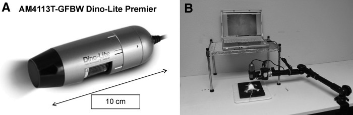
Materials and methods: Patient colon tumors were initially established subcutaneously in nonobese diabetic (NOD)/severe combined immune deficiency (SCID) mice immediately after surgery. The tumors were then harvested from NOD/SCID mice and passed orthotopically in nude mice to make patient-derived orthotopic xenograft (PDOX) models. Eight weeks after orthotopic implantation, a monoclonal anti-carcinoembryonic antigen (CEA) antibody conjugated with AlexaFluor 488 (Molecular Probes Inc., Eugene, OR) was delivered to the PDOX models as a single intravenous dose 24 hours before laparotomy. A hand-held portable fluorescence imaging device was used.
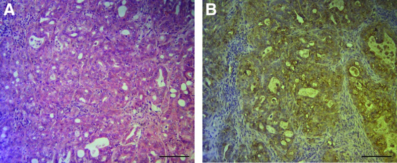
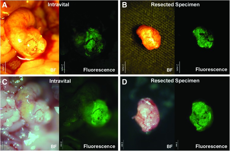
Results: The primary tumor was clearly visible at laparotomy with the portable fluorescence imaging system. Frozen section microscopy of the resected specimen demonstrated that the anti-CEA antibody selectively labeled cancer cells in the colon cancer PDOX. The tumor was completely resected under fluorescence navigation. Histologic evaluation of the resected specimen demonstrated that cancer cells were not present in the margins, indicating successful tumor resection. The FGS animals remained tumor free for over 6 months.
Conclusions: The results of the present report indicate that FGS using a fluorophore-conjugated anti-CEA antibody and portable imaging system improves efficacy of resection for CEA-positive colorectal cancer. These data provide the basis for clinical trials.
Figures


References
-
- Bouvet M, Hoffman RM. Glowing tumors make for better detection and resection. Sci Transl Med 2011;3(110):110fs110 - PubMed
-
- Ruo L, Guillem JG. Surgical management of primary colorectal cancer. Surg Oncol 1998;7:153–163 - PubMed
-
- Fujiwara T, Kagawa S, Kishimoto H, et al. Enhanced antitumor efficacy of telomerase-selective oncolytic adenoviral agent OBP-401 with docetaxel: Preclinical evaluation of chemovirotherapy. Int J Cancer 2006;119:432–440 - PubMed
-
- Kishimoto H, Kojima T, Watanabe Y, et al. In vivo imaging of lymph node metastasis with telomerase-specific replication-selective adenovirus. Nat Med 2006;12:1213–1219 - PubMed
Publication types
MeSH terms
Substances
Grants and funding
LinkOut - more resources
Full Text Sources
Other Literature Sources
