

Flow cytometric characterization and clinical outcome of CD4+ T-cell lymphoma in dogs: 67 cases
- PMID: 24495161
- PMCID: PMC4857986
- DOI: 10.1111/jvim.12304
Flow cytometric characterization and clinical outcome of CD4+ T-cell lymphoma in dogs: 67 cases
Erratum in
- J Vet Intern Med. 2014 May-Jun;28(3):1139
Abstract
Background: Canine T-cell lymphoma (TCL) is conventionally considered an aggressive disease, but some forms are histologically and clinically indolent. CD4 TCL is reported to be the most common subtype of TCL. We assessed flow cytometric characteristics, histologic features when available, and clinical outcomes of CD4+ TCL to determine if flow cytometry can be used to subclassify this group of lymphomas.
Objective: To test the hypothesis that canine CD4+ T-cell lymphoma (TCL) is a homogeneous group of lymphomas with an aggressive clinical course.
Animals: Sixty-seven dogs diagnosed with CD4+ TCL by flow cytometry and treated at 1 of 3 oncology referral clinics.
Methods: Retrospective multivariable analysis of outcome in canine CD4+ TCL including patient characteristics, treatment, and flow cytometric features.
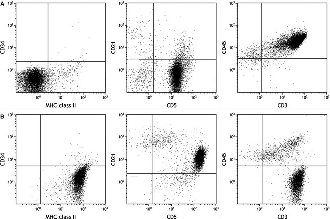
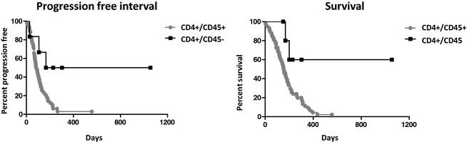
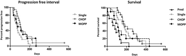
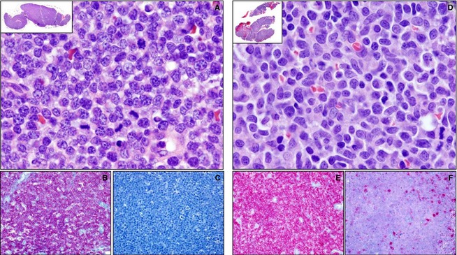
Results: The majority of CD4+ TCL were CD45+, expressed low class II MHC, and exhibited an aggressive clinical course independent of treatment regimen (median survival, 159 days). Histologically, CD4+ TCL were classified as lymphoblastic or peripheral T cell. Size of the neoplastic lymphocytes had a modest effect on both PFI and survival in this group. A small number of CD4+ TCL were CD45- and class II MHC high, and exhibited an apparently more indolent clinical course (median survival not yet reached).
Conclusions and clinical importance: Although the majority of CD4+ TCL in dogs had uniform clinical and flow cytometric features and an aggressive clinical course, a subset had a unique immunophenotype that predicts significantly longer survival. This finding strengthens the utility of flow cytometry to aid in the stratification of canine lymphoma.
Keywords: Canine; Immunophenotyping; Lymphoblastic; Peripheral T cell; Prognosis.
Copyright © 2014 by the American College of Veterinary Internal Medicine.
Figures
References
-
- Ponce F, Magnol JP, Ledieu D, et al. Prognostic significance of morphological subtypes in canine malignant lymphomas during chemotherapy. Vet J 2004;167:158–166. - PubMed
-
- Valli VE, Vernau W, de Lorimier L‐P, et al. Canine indolent nodular lymphoma. Vet Pathol 2006;43:241–256. - PubMed
-
- Sozmen M, Tasca S, Carli E, et al. Use of fine needle aspirates and flow cytometry for the diagnosis, classification, and immunophenotyping of canine lymphomas. J Vet Diagn Invest 2005;17:323–330. - PubMed
-
- Gelain ME, Mazzilli M, Riondato F, et al. Aberrant phenotypes and quantitative antigen expression in different subtypes of canine lymphoma by flow cytometry. Vet Immunol Immunopathol 2008;121:179–188. - PubMed
-
- Lurie DM, Milner RJ, Suter SE, et al. Immunophenotypic and cytomorphologic subclassification of T‐cell lymphoma in the Boxer breed. Vet Immunol Immunopathol 2008;125:102–110. - PubMed
MeSH terms
Substances
LinkOut - more resources
Full Text Sources
Other Literature Sources
Research Materials
Miscellaneous