

ADAM17 mediates OSCC development in an orthotopic murine model
- PMID: 24495306
- PMCID: PMC3928084
- DOI: 10.1186/1476-4598-13-24
ADAM17 mediates OSCC development in an orthotopic murine model
Abstract
Background: ADAM17 is one of the main sheddases of the cells and it is responsible for the cleavage and the release of ectodomains of important signaling molecules, such as EGFR ligands. Despite the known crosstalk between ADAM17 and EGFR, which has been considered a promising targeted therapy in oral squamous cell carcinoma (OSCC), the role of ADAM17 in OSCC development is not clear.
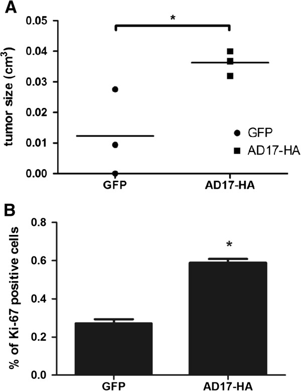
Method: In this study the effect of overexpressing ADAM17 in cell migration, viability, adhesion and proliferation was comprehensively appraised in vitro. In addition, the tumor size, tumor proliferative activity, tumor collagenase activity and MS-based proteomics of tumor tissues have been evaluated by injecting tumorigenic squamous carcinoma cells (SCC-9) overexpressing ADAM17 in immunodeficient mice.
Results: The proteomic analysis has effectively identified a total of 2,194 proteins in control and tumor tissues. Among these, 110 proteins have been down-regulated and 90 have been up-regulated in tumor tissues. Biological network analysis has uncovered that overexpression of ADAM17 regulates Erk pathway in OSCC and further indicates proteins regulated by the overexpression of ADAM17 in the respective pathway. These results are also supported by the evidences of higher viability, migration, adhesion and proliferation in SCC-9 or A431 cells in vitro along with the increase of tumor size and proliferative activity and higher tissue collagenase activity as an outcome of ADAM17 overexpression.
Conclusion: These findings contribute to understand the role of ADAM17 in oral cancer development and as a potential therapeutic target in oral cancer. In addition, our study also provides the basis for the development of novel and refined OSCC-targeting approaches.
Figures


Similar articles
-
MicroRNA-224, negatively regulated by c-jun, inhibits growth and epithelial-to-mesenchymal transition phenotype via targeting ADAM17 in oral squamous cell carcinoma.J Cell Mol Med. 2019 Aug;23(8):4913-4920. doi: 10.1111/jcmm.14107. Epub 2019 Jun 17. J Cell Mol Med. 2019. PMID: 31207072 Free PMC article.
-
Increase of disintergin metalloprotease 10 (ADAM10) expression in oral squamous cell carcinoma.Cancer Lett. 2007 Jan 8;245(1-2):33-43. doi: 10.1016/j.canlet.2005.10.019. Epub 2005 Nov 23. Cancer Lett. 2007. PMID: 16309826
-
MicroRNA-186 serves as a tumor suppressor in oral squamous cell carcinoma by negatively regulating the protein tyrosine phosphatase SHP2 expression.Arch Oral Biol. 2018 May;89:20-25. doi: 10.1016/j.archoralbio.2018.01.016. Epub 2018 Jan 31. Arch Oral Biol. 2018. PMID: 29407635
-
Overexpression of angiopoietin 2 promotes the formation of oral squamous cell carcinoma by increasing epithelial-mesenchymal transition-induced angiogenesis.Cancer Gene Ther. 2016 Sep;23(9):295-302. doi: 10.1038/cgt.2016.30. Epub 2016 Aug 5. Cancer Gene Ther. 2016. PMID: 27492854 Free PMC article.
-
[Effect of circular RNA hsa_circ_0008898 on oral squamous cell carcinoma and its mechanism].Zhonghua Kou Qiang Yi Xue Za Zhi. 2020 Aug 9;55(8):578-585. doi: 10.3760/cma.j.cn112144-20200109-00006. Zhonghua Kou Qiang Yi Xue Za Zhi. 2020. PMID: 32842350 Chinese.
Cited by
-
Quantitative proteomic analysis for novel biomarkers of buccal squamous cell carcinoma arising in background of oral submucous fibrosis.BMC Cancer. 2016 Aug 2;16:584. doi: 10.1186/s12885-016-2650-1. BMC Cancer. 2016. PMID: 27485544 Free PMC article.
-
iRhom2 in the pathogenesis of oral squamous cell carcinoma.Mol Biol Rep. 2020 May;47(5):3987-3992. doi: 10.1007/s11033-020-05381-y. Epub 2020 Mar 31. Mol Biol Rep. 2020. PMID: 32236893 Free PMC article.
-
MicroRNA-224, negatively regulated by c-jun, inhibits growth and epithelial-to-mesenchymal transition phenotype via targeting ADAM17 in oral squamous cell carcinoma.J Cell Mol Med. 2019 Aug;23(8):4913-4920. doi: 10.1111/jcmm.14107. Epub 2019 Jun 17. J Cell Mol Med. 2019. PMID: 31207072 Free PMC article.
-
Repurposing EGFR Inhibitors for Oral Cancer Pain and Opioid Tolerance.Pharmaceuticals (Basel). 2023 Nov 3;16(11):1558. doi: 10.3390/ph16111558. Pharmaceuticals (Basel). 2023. PMID: 38004424 Free PMC article. Review.
-
Multifactorial Contribution of Notch Signaling in Head and Neck Squamous Cell Carcinoma.Int J Mol Sci. 2019 Mar 26;20(6):1520. doi: 10.3390/ijms20061520. Int J Mol Sci. 2019. PMID: 30917608 Free PMC article. Review.
References
-
- Murphy G. The ADAMs: signalling scissors in the tumour microenvironment. Nat Rev Cancer. 2008;8:929–941. - PubMed
Publication types
MeSH terms
Substances
LinkOut - more resources
Full Text Sources
Other Literature Sources
Medical
Molecular Biology Databases
Research Materials
Miscellaneous
