. 2014 Feb 5:11:28.
doi: 10.1186/1742-2094-11-28.

Suppression of neuroinflammation in forebrain-specific Cdk5 conditional knockout mice by PPARγ agonist improves neuronal loss and early lethality

Affiliations

Suppression of neuroinflammation in forebrain-specific Cdk5 conditional knockout mice by PPARγ agonist improves neuronal loss and early lethality

Elias Utreras et al. J Neuroinflammation. .

Abstract

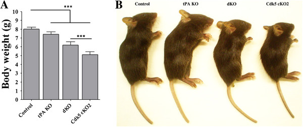
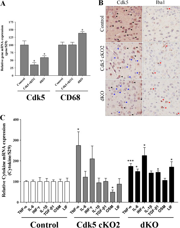
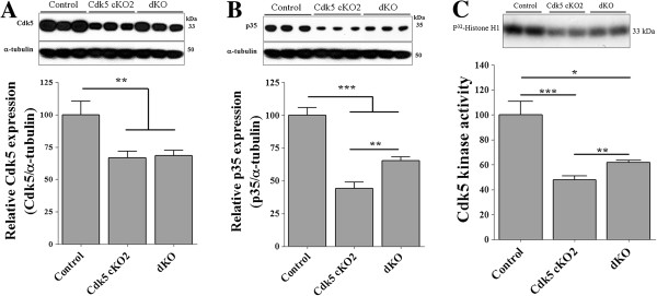
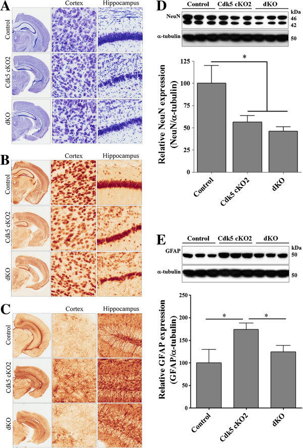
Background: Cyclin-dependent kinase 5 (Cdk5) is essential for brain development and function, and its deregulated expression is implicated in some of neurodegenerative diseases. We reported earlier that the forebrain-specific Cdk5 conditional knockout (cKO) mice displayed an early lethality associated with neuroinflammation, increased expression of the neuronal tissue-type plasminogen activator (tPA), and neuronal migration defects.

Methods: In order to suppress neuroinflammation in the cKO mice, we first treated these mice with pioglitazone, a PPARγ agonist, and analyzed its effects on neuronal loss and longevity. In a second approach, to delineate the precise role of tPA in neuroinflammation in these mice, we generated Cdk5 cKO; tPA double knockout (dKO) mice.

Results: We found that pioglitazone treatment significantly reduced astrogliosis, microgliosis, neuronal loss and behavioral deficit in Cdk5 cKO mice. Interestingly, the dKO mice displayed a partial reversal in astrogliosis, but they still died at early age, suggesting that the increased expression of tPA in the cKO mice does not contribute significantly to the pathological process leading to neuroinflammation, neuronal loss and early lethality.

Conclusion: The suppression of neuroinflammation in Cdk5 cKO mice ameliorates gliosis and neuronal loss, thus suggesting the potential beneficial effects of the PPARγ agonist pioglitazone for the treatment for neurodegenerative diseases.


Figures

Figure 1
Figure 1
PPARγ agonist pioglitazone suppressed inflammation and partially reversed neuronal loss in Cdk5 cKO1 mice. (A) Survival curves for untreated and treated Cdk5 cKO1 mice from P30 to P60. Survival rate for treated mice (n = 16) is better than that of untreated mice (n = 13). (B) Average body weights of untreated (n = 3) and treated (n = 3) male Cdk5 cKO1 mice and littermate male Cdk5f/f (control) mice (n = 3) at P60. *P < 0.05. (C), (D) Immunohistochemical (IHC) analyses of control, untreated, and treated Cdk5 cKO1 mice in cerebral cortex (C) and hippocampus (D) at P60. Coronal sections from these mice were stained with antibodies for NeuN, GFAP and Iba1. Scale bar = 100 μm. (E-G) Bar graphs indicate average cell densities of neurons (E), activated astrocytes (F) and microglia cells (G) in control, untreated and treated Cdk5 cKO1 mice at P60. ***P < 0.001 between control versus Cdk5 cKO1. †P < 0.05; ‡P < 0.01; §P < 0.001 between no treated versus treated (t-test).
Figure 2
Figure 2
Generation of Cdk5 cKO2; tPA KO (dKO) mice. (A) Body weights of 17-day-old mice of four different genotypes. (B) Representative images of control, tPA KO, Cdk5 cKO2 and dKO mice. All data are presented as the mean and SEM (n = 6 to 12 mice per group), ***P < 0.001 (t-test).
Figure 3
Figure 3
Increased microgliosis and pro-inflammatory cytokines in the brain of dKO mice. (A) qPCR analysis of Cdk5 and CD68 mRNA levels (a marker of activated microglia) normalized against S29. Total RNA was obtained from brain cortex of control, Cdk5 cKO2 and dKO mice at P17. There was a decreased Cdk5 mRNA levels on Cdk5 cKO2 and dKO mice. In contrast, CD68 was increased only on dKO mice as compared with control mice. (B) Right panel shows a representative IHC against Cdk5 and left panel an IHC against Iba1 of cortex coronal section from control, Cdk5 cKO2 and dKO mice at P17. Decreased Cdk5 expression was observed in the cortex of Cdk5 cKO2 and dKO mice (Blue head arrows as compared with black head arrows). IHC Iba1 shows that Iba1 positive cells are few in the cortex of the control mice as compared with Cdk5 cKO2 and dKO mice (red head arrows). Also, Iba1 positive cells are more apparent on dKO as compared with Cdk5 cKO2 mice. (C) Real-time PCR analysis of mRNA levels of several pro-inflammatory cytokines normalized against S29. All data are presented as the mean and SEM (n = 3 to 5). *P < 0.05; ***P < 0.001 (t-test).
Figure 4
Figure 4
Decreased Cdk5 and p35 protein expression and Cdk5 activity in the brain of Cdk5 cKO2 and dKO mice. (A) Representative Western blot analysis showing Cdk5 protein levels in brain cortex of control, Cdk5 cKO2 and dKO mice at P17. Lower panel showed a quantification of Western blots for Cdk5. α-tubulin was used as loading control. (B) Representative Western blot analysis showing p35 protein levels in brain cortex of control, Cdk5 cKO2 and dKO mice at P17. Lower panel showed a quantification of Western blots of p35. α-tubulin was used as loading control. (C) Cdk5 kinase activity was measured from immunoprecipitates of proteins obtained from brain cortex of control, Cdk5 cKO2 and dKO mice at P17. All data are presented as the mean and SEM (n = 3 to 12). *P < 0.05, **P < 0.01, ***P < 0.001 (t-test).
Figure 5
Figure 5
Increased neuronal loss and partial rescue of astrogliosis in the brain of dKO mice. (A) Nissl staining of coronal brain sections of control, Cdk5 cKO2 and dKO mice at P17. In higher magnification we observed a decreased of the number of cells on cortex and hippocampus of Cdk5 cKO2 and dKO mice. (B) Immunohistochemistry (IHC) against NeuN, a marker of neurons, of coronal brain sections of control, Cdk5 cKO2 and dKO mice at P17. In higher magnification we observed a decreased of the number of NeuN positive cells on cortex and hippocampus of Cdk5 cKO2 and dKO mice. (C) IHC against GFAP, a marker of astrogliosis, of coronal brain sections of control, Cdk5 cKO2 and dKO mice at P17. In higher magnification we observed an increased number of GFAP positive cells on cortex and hippocampus of Cdk5 cKO2. In contrast, there were a decreased number of GFAP positive cells on the same areas of brain of dKO mice. (D) Representative Western blot analysis showing NeuN protein levels in brain cortex of control, Cdk5 cKO2 and dKO mice at P17. Lower panel showed a quantification of Western blots of NeuN. α-tubulin was used as loading control. (E) Representative Western blot analysis showing GFAP protein levels in brain cortex of control, Cdk5 cKO2 and dKO mice at P17. Lower panel showed a quantification of Western blots of GFAP. α-tubulin was used as loading control. All data are presented as the mean and SEM (n = 3 to 6). *P < 0.05 (t-test).

References

    1. Dhavan R, Tsai LH. A decade of CDK5. Nat Rev Mol Cell Biol. 2001;2:749–759. doi: 10.1038/35096019. - DOI - PubMed
    1. Lew J, Huang QQ, Qi Z, Winkfein RJ, Aebersold R, Hunt T, Wang JH. A brain-specific activator of cyclin-dependent kinase 5. Nature. 1994;371:423–426. doi: 10.1038/371423a0. - DOI - PubMed
    1. Dhariwala FA, Rajadhyaksha MS. An unusual member of the Cdk family: Cdk5. Cell Mol Neurobiol. 2008;28:351–369. doi: 10.1007/s10571-007-9242-1. - DOI - PMC - PubMed
    1. Borquez DA, Olmos C, Alvarez S, Di Genova A, Maass A, Gonzalez-Billault C. Bioinformatic survey for new physiological substrates of Cyclin-dependent kinase 5. Genomics. 2013;101:221–228. doi: 10.1016/j.ygeno.2013.01.003. - DOI - PubMed
    1. Contreras-Vallejos E, Utreras E, Gonzalez-Billault C. Going out of the brain: non-nervous system physiological and pathological functions of Cdk5. Cell Signal. 2012;24:44–52. doi: 10.1016/j.cellsig.2011.08.022. - DOI - PubMed

Publication types

MeSH terms

LinkOut - more resources