Review
. 2014 Apr:133:92-103.
doi: 10.1016/j.jinorgbio.2014.01.005. Epub 2014 Jan 21.

Modulation of nuclear receptor function by cellular redox poise

Affiliations
Review

Modulation of nuclear receptor function by cellular redox poise

Eric L Carter et al. J Inorg Biochem. 2014 Apr.

Abstract

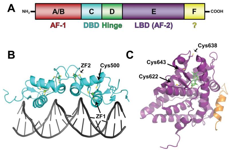
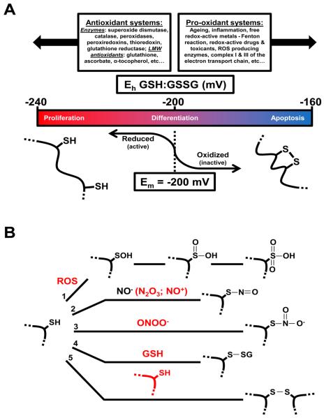
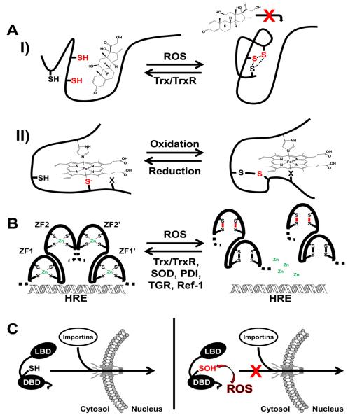
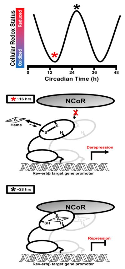
Nuclear receptors (NRs) are ligand-responsive transcription factors involved in diverse cellular processes ranging from metabolism to circadian rhythms. This review focuses on NRs that contain redox-active thiol groups, a common feature within the superfamily. We will begin by describing NRs, how they regulate various cellular processes and how binding ligands, corepressors and/or coactivators modulate their activity. We will then describe the general area of redox regulation, especially as it pertains to thiol-disulfide interconversion and the cellular systems that respond to and govern this redox equilibrium. Lastly, we will discuss specific examples of NRs whose activities are regulated by redox-active thiols. Glucocorticoid, estrogen, and the heme-responsive receptor, Rev-erb, will be described in the most detail as they exhibit archetypal redox regulatory mechanisms.

Keywords: Hormone; Nuclear receptor; Oxidative stress; Redox; Thiol–disulfide; Transcription.


Figures

Fig. 1
Fig. 1. Nuclear receptor domain organization and three-dimensional structure using glucocorticoid receptor as an example
(A) Modular domain organization of a typical NR. The N-terminal A/B region harbors AF-1 ligand-independent regulatory activity and is a site of phosphorylation. A/B is followed by the highly conserved C region, or DBD that is connected to the LBD (E region, containing ligand-dependent AF-2 activity) by the hinge (D region). Some receptors contain a F region with unknown function. (B) Three-dimensional structure of the rat GR DBD homodimer in complex with a palindromic GRE where the two half-sites are separated by four base pairs (PDB access code 1GLU). Cysteine sulfhydryls involved in redox modulation are highlighted in yellow. In each subunit of the dimer, eight out of nine redox-active cysteine sulfhydryls comprise the two Cys4 zinc-finger motifs (ZF1 and ZF2). The last sulfhydryl of Cys500 (equivalent to Cys481 in the human GR sequence) resides in NLS1 and is conserved among nuclear receptors. (C) Three-dimensional structure of the human GR LBD in complex with the agonist dexamethasone (depicted as sticks with carbon atoms in green, oxygen in red, and fluorine in cyan) and a peptide derived from the coactivator TIF2 (orange) harboring a LXXLL motif (PDB access code 1M2Z). The three cysteine sulfhydryls implicated in redox modulation of ligand binding are highlighted in yellow and labeled; Cys638 and Cys643 are the pair of arsenite-reactive proximal thiols.
Fig. 2
Fig. 2. Cellular redox systems interface with protein thiols
(A) Redox homeostasis is maintained as a balance between opposing antioxidant and pro-oxidant systems. However, homeostasis is a relative term considering the ambient potential of glutathione, the major cellular redox buffer, can vary dramatically depending on the growth stage of a cell. Proteins containing redox active thiol-disulfide couples will respond to the ambient potential in a Nernstian fashion as long as the Em (midpoint potential) of the couple falls within the physiologically relevant range. To illustrate this point a hypothetical thiol-disulfide that controls the activity of a protein is depicted; the Em of the couple is −200 mV and the dithiol form of the protein is active while the disulfide is inactive. When the cell is proliferating and the ambient potential is highly reducing the couple exists as a dithiol. Conversely, a cell undergoing apoptosis can be +80 mV oxidized favoring an intramolecular disulfide that alters quaternary structure and inactivates the protein. Adapted from [9]. (B) Many other thiol oxidation products exist besides intramolecular disulfides. For simplicity a single reduced protein thiol is depicted with oxidizing species in red: 1) ROS can successively oxidize thiols to sulfenic (-SOH), sulfinic (-SO2H), and sulfonic (-SO3H) acids; 2) Autooxidation products of nitric oxide, such as dinitrogen trioxide, or species with NO+ character react with thiols to form thionitrites (S-nitrosothiols); 3) Peroxynitrite reacts with thiols generating a thionitrate that may be an intermediate in the formation of disulfides, a major peroxynitrite-driven thiol oxidation product [182]; 4) Low molecular weight thiols like GSH can form disulfides with protein thiols under oxidizing conditions; 5) Similarly, thiols of different proteins can form intermolecular disulfides. Adapted from [183].
Fig. 3
Fig. 3. Key modes of redox modulation of nuclear receptor function
(A) Two examples are depicted in which the redox status of sulfhydryls in the ligand-binding domain govern the ability of the receptor to effectively associate with ligand. I) In the thiol-reduced state the glucocorticoid receptor binds the agonist, dexamethasone that ultimately leads to nuclear import and the regulation of target gene transcription. Under conditions of oxidative stress ROS cause the formation of disulfide bonds between any of three cysteine sulfhydryls in close proximity to the ligand-binding pocket. Inspection of the GR LBD crystal structure suggests that in order for these disulfides to form a large conformational change would have to take place that could occlude access to dexamethasone or other glucocorticoids. The dashed lines represent potential disulfides and the two red sulfhydryls comprise the arsenite-reactive proximal thiols unique to GR. In the reverse direction, Trx and TrxR have been implicated in the reduction of thiol oxidized GR and the recovery of steroid-binding capacity. II) Under reducing conditions the Rev-erbβ LBD binds ferric heme with high affinity as a 6-coordinate complex with histidine and cysteine thiolate (depicted in red, Cys384) axial ligands. An oxidizing environment leads to the sequestration of the axial cysteine thiolate as a disulfide with Cys374 and the concomitant replacement of Cys384 as a heme ligand by an unidentified neutral residue, X. The thiol-oxidized protein has a ~5-fold decreased affinity for heme which could have a significant impact on the ability of Rev-erbβ to recruit NCoR and repress target gene transcription. (B) NR DBD zinc-fingers are targets of ROS damage. Under reducing conditions zinc-fingers are maintained as a tetrahedral complex between cysteine thiolates and Zn2+ that impart structural stability important for binding DNA and dimerization. ROS lead to the formation of disulfides between zinc-finger sulfhydryls with subsequent ejection of Zn2+ and structural destabilization that ultimately causes dimer dissociation and a loss of DNA binding capacity. As demonstrated for ER, ZF2 is highly susceptible to thiol oxidation thus disulfide bonds in ZF2 are shown in red. The reduction and recovery of oxidized ER DBD disulfides are facilitated by an interaction with redox mediators in the nucleus including Trx/TrxR, SOD, PDI, and Ref-1. Trx has also been implicated in the repair of thiol oxidized GR DBD, the hybrid thiol-disulfide oxidoreductase TGR similarly affects RAR, and Ref-1 modulates TR activity suggesting a general mechanism in which thiol-disulfide oxidoreductases, chaperones, and antioxidant enzymes maintain the reduced and active forms of NRs in the cell. HRE: Hormone Response Element. (C) Nuclear translocation of GR is subject to redox modulation through the oxidation of Cys500 (rat amino acid numbering, equivalent to Cys481 in human GR), a conserved cysteine sulfhydryl that resides in NLS1 C-terminal to ZF2 (depicted as a –SH group). Under reducing conditions importin proteins interact with NLS1 leading to the translocation of GR to the nucleus through a nuclear pore. On the other hand, ROS that arise during oxidative stress can oxidize Cys500 and impede nuclear translocation. A sulfenic acid intermediate is depicted as it is unclear what the Cys500 oxidation product is, but could certainly include an intra- or intermolecular disulfide, or higher sulfur oxide that may inhibit the ability of GR to interact with importin proteins.
Fig. 4
Fig. 4. A working hypothesis for the role of the Rev-erbβ thiol-disulfide switch in syncing repressor function with rhythmic changes in suprachiasmatic nucleus cellular redox status
Several redox systems display rhythmic cycles of reduction/oxidation in rodent SCN tissue including FAD/NADPH cofactors, protein glutathionylation, and ascorbate [154]. In terms of rodent circadian time, SCN tissue redox status (measured by the ratio of FAD:NADPH) is maximally oxidized at ~16 h (early night); these conditions would seemingly favor the disulfide form of the Rev-erbβ thiol-disulfide switch, lowering the affinity of the receptor for heme and impeding the ligand-dependent recruitment of NCoR ultimately leading to the derepression of target gene transcription. Conversely, at ~28 hours clock time (daytime) SCN tissue is maximally reduced and would favor the dithiol form of the switch which has a high affinity for heme with His (H) and Cys axial ligands. In the heme bound state Rev-erbβ recruits NCoR leading to gene repression.

Similar articles

Cited by

References

    1. Burris TP, Solt LA, Wang Y, Crumbley C, Banerjee S, Griffett K, Lundasen T, Hughes T, Kojetin DJ. Pharmacol. Rev. 2013;65:710–778. - PMC - PubMed
    1. Moore JT, Collins JL, Pearce KH. ChemMedChem. 2006;1:504–523. - PubMed
    1. Kumar V, Green S, Stack G, Berry M, Jin J-R, Chambon P. Cell. 1987;51:941–951. - PubMed
    1. Tsai M, O'Malley BW. Annu. Rev. Biochem. 1994;63:451–486. - PubMed
    1. Aranda A, Pascual A. Physiol. Rev. 2001;81:1269–1304. - PubMed

Publication types

LinkOut - more resources