. 2014 Apr;196(4):1091-102.
doi: 10.1534/genetics.113.156323. Epub 2014 Feb 4.

The extracellular matrix protein artichoke is required for integrity of ciliated mechanosensory and chemosensory organs in Drosophila embryos

Affiliations

The extracellular matrix protein artichoke is required for integrity of ciliated mechanosensory and chemosensory organs in Drosophila embryos

Marta Andrés et al. Genetics. 2014 Apr.

Abstract

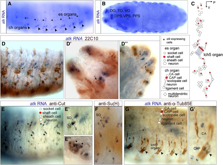
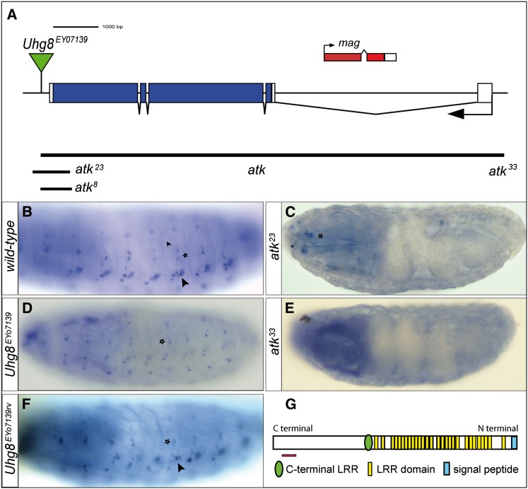
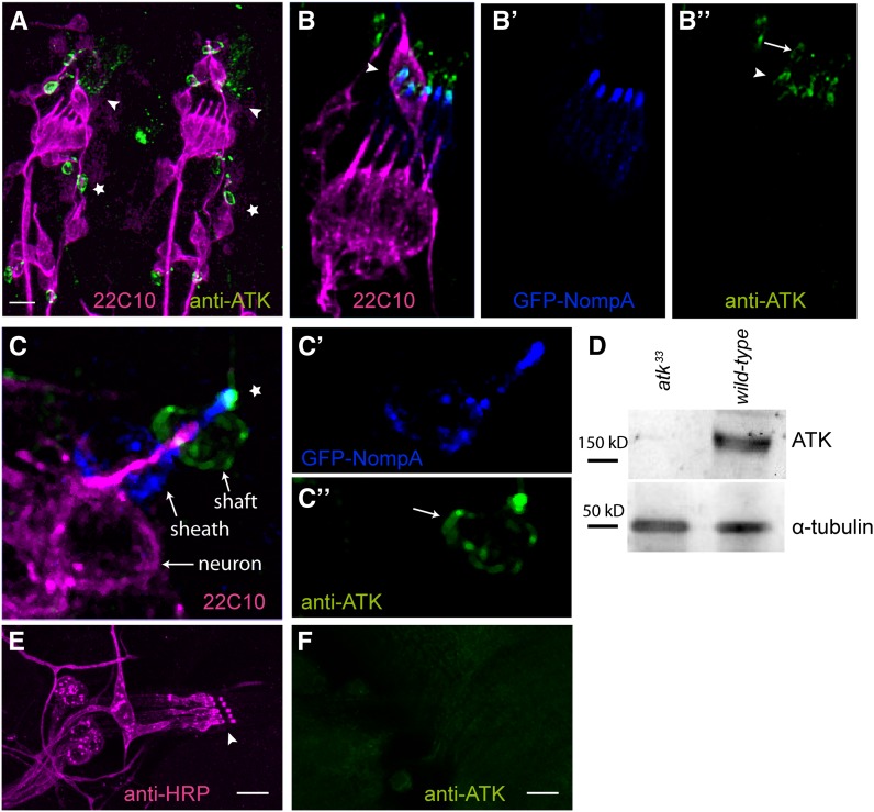
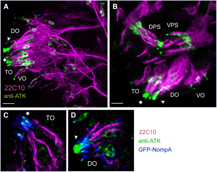
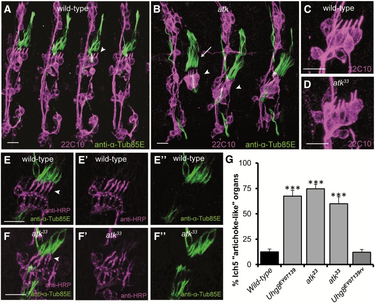
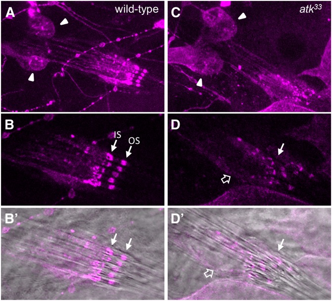
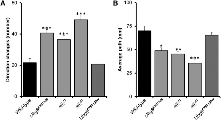
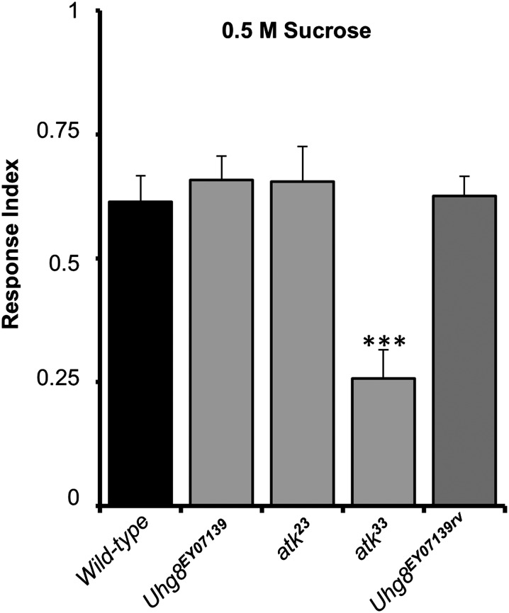
Sensory cilia are often encapsulated by an extracellular matrix (ECM). In Caenorhabditis elegans, Drosophila melanogaster, and vertebrates, this ECM is thought to be directly involved in ciliary mechanosensing by coupling external forces to the ciliary membrane. Drosophila mechano- and chemosensory cilia are both associated with an ECM, indicating that the ECM may have additional roles that go beyond mechanosensory cilium function. Here, we identify Artichoke (ATK), an evolutionarily conserved leucine-rich repeat ECM protein that is required for normal morphogenesis and function of ciliated sensilla in Drosophila. atk is transiently expressed in accessory cells in all ciliated sensory organs during their late embryonic development. Antibody stainings show ATK protein in the ECM that surrounds sensory cilia. Loss of ATK protein in atk null mutants leads to cilium deformation and disorientation in chordotonal organs, apparently without uncoupling the cilia from the ECM, and consequently to locomotion defects. Moreover, impaired chemotaxis in atk mutant larvae suggests that, based on ATK protein localization, the ECM is also crucial for the correct assembly of chemosensory receptors. In addition to defining a novel ECM component, our findings show the importance of ECM integrity for the proper morphogenesis of ciliated organs in different sensory modalities.

Keywords: LRR proteins; chordotonal organ; dendritic cap; extracellular matrix; sensory cilium.


Figures

Figure 1
Figure 1
atk is expressed by an accessory cell in every ciliated sensory organ. (A and B) atk in situ hybridization in wild-type stage 16 embryos. (A) Lateral view showing atk in situ signal in a pattern consistent with expression in ch organs (arrowheads) and es organs (stars). (B) Horizontal view showing atk expression in chemosensory organs: dorsal organ (DO), terminal organ (TO), ventral organ (VO), dorsal, ventral and posterior pharyngeal organs (DPS, VPS, and PPS, respectively). (C) Schematic diagram of mechanosensory organs per hemisegment showing atk-expressing cells (red). (D–G) Embryos double-stained for atk in situ (blue signal) and different primary antibodies (brown signal). (D) atk is not expressed by ciliated neurons labeled by the neuronal marker mAb 22C10 (arrows, D–D′′). (E and F) In es and chemosensory organs, atk is expressed by the shaft cell: it colocalizes with anti-Cut signal (stars, E–E′′), which labels socket (SO) and shaft (S) cells, but does not colocalize with anti-Su(H), which labels the socket (F). (G) In ch organs, atk is expressed by the cap cell (CAP) based on atk-expressing cell position and colocalization with anti-α-Tub85E (arrowheads, G and G′). CA: cap attachment cell; N: neuron.
Figure 2
Figure 2
Generation of atk mutant alleles and anti-ATK antibody. (A) Schematic of atk locus. Shown are the location of the P{EPgy2} element Uhg8EY07139, extent of deletion in the alleles generated—hypomorphs atk8 and atk23 and null allele atk33— and location of the gene mag. (B–F) Stage 16 embryos hybridized with an atk antisense RNA probe. (B) Wild-type embryo showing hybridization in es organs (star) and ch organs (arrowhead). (C) atk23 mutant embryos lack atk expression in mechanosensory organs, but expression persists in chemosensory organs (asterisk). (D) Embryos homozygous for Uhg8EY07139 lose atk expression in ch organs (arrowhead in B and F) but keep it in es organs (star). (E) atk33 homozygous embryos show no atk expression. (F) Uhg8EY07139rv show normal atk expression. (G) Predicted ATK protein with 29 LRRs. Red line indicates the region against which anti-ATK antibody was raised.
Figure 3
Figure 3
ATK localizes to the distal region of the dendritic cap in late embryonic mechanosensory organs. (A) Stage 16 embryonic hemisegments immunostained with the neuronal marker mAb22C10 (magenta) and with anti-ATK (green). ATK localizes apically to the sensory cilia in ch (arrowhead) and es (star) organs. (B and C) Detailed view of embryonic mechanosensory organs additionally showing GFP-NOMPA (blue) in the dendritic cap. Single channel images are shown for GFP-NOMPA (B′ and C′) and anti-ATK (B″ and C″). In lch5 (B) and es (C) organs, ATK localizes to the distal region of the dendritic cap (arrowhead in B, star in C), where it partially overlaps with GFP-NOMPA. The cytoplasm of cap and shaft cells are also stained (arrows). (D) Western blot with anti-ATK antibody. (E and F) ATK protein is not present in larval mechanosensory organs. Second instar larval lch5 organ stained with anti-HRP (E) shows no detectable anti-ATK staining. (F) Bars, 10 µm.
Figure 4
Figure 4
ATK localizes to a supporting ECM in late embryonic chemosensory organs. (A and B) Stage 16 embryonic chemosensory organs immunostained with the neuronal marker mAb22C10 (magenta) and anti-ATK (green). DO: dorsal organ; TO: terminal organ; VO: ventral organ; DPS: dorsal pharyngeal organ; VPS: ventral pharyngeal organ. In DO (arrowhead) and TO (star), ATK localizes to the tip of the chemosensory cilia. Cell bodies of atk-expressing cells are also stained. Bars, 10 µm. (C and D) Detailed view of TO (C) and DO (D) additionally showing GFP-NOMPA (blue). (C) In TO, ATK localizes at the tip of the GFP-NOMPA region (star), where gustatory neurons are exposed to the environment. (D) In DO, ATK forms a vacuole-like structure in the center of the cilia cluster (arrowhead).
Figure 5
Figure 5
atk mutants show abnormal embryonic lch5 organ morphology. (A–D) Stage 16 embryonic hemisegments stained with neuronal marker mAb22C10 (magenta, labeling cell body and inner dendritic segment) and anti-α-Tub85E (green), which labels all ch organ accessory cells except the scolopale cell. (A and C) In wild-type lch5 organs, sensory cilia are parallel and point in the same direction (arrowhead). Neuronal cell bodies are aligned in a row. (B and D) In atk33 embryos, many lch5 organs show an artichoke-like phenotype with nonparallel dendrites and misaligned cell bodies (arrowheads in B). Cap cells also show defects (arrow in B). (E and F) Lch5 organs of stage 16 embryos, stained with anti-HRP (magenta, E′ and F′) that labels the entire cilium, and anti-α-Tub85E (green, E″ and F″). The cilium tip contacts the dendritic cap in both wild-type (E, arrowhead) and atk33 mutants (F, arrowhead). Bars, 10 µm. (G) Percentage of artichoke-like lch5 organs per hemisegment and genotype. Embryos were stained with mAb22C10 to label the sensory neurons and scored blind to genotype. Significant difference was established by the Kruskal–Wallis test followed by a nonparametric post-hoc test to compare each genotype with wild type. ***P < 0.001.
Figure 6
Figure 6
atk mutant larvae show abnormal lch5 organ morphology and altered anti-HRP signal in cilia. Third instar larval lch5 organs stained with anti-HRP (magenta) in control (A and B) and atk33 mutants (C and D). (A and C) General view of neuronal cell bodies (arrowheads) and cilia. (B and D) High magnification of cilia showing anti-HRP signal, alone (B and D) or merged with a bright-field image (B′ and D′). In control lh5 organs (B), cilia are parallel and aligned with anti-HRP signal concentrated in two bands corresponding to inner (IS) and outer (OS) segments (Ma and Jarman 2011). In atk mutants (D), alignment of cilia is lost (open arrow points to a cilium that is bent and separated from the others), and distribution of anti-HRP immunoreactivity is severely disrupted.
Figure 7
Figure 7
atk mutants show larval locomotion defects. Locomotion was scored for 100 sec by measuring the total number of direction changes (A) and the average path length (mm) (B). Means are represented. Error bars indicate SEM. Significant difference with respect to wild type was established by Bonferroni-corrected t-test. *P < 0.05; ** P < 0.01; ***P < 0.005.
Figure 8
Figure 8
atk mutant larvae show a significantly reduced response to sucrose. Response index for election between 1% agarose and 1% agarose + 0.5 M sucrose. atk33 null mutants show defects in chemosensation. atk23 hypomorphs and Uhg8EY07139 homozygous larvae, which keep atk expression in chemosensory organs, do not show defects. Therefore, ATK seems to be required for chemosensation, although an effect of additional genes deleted by the atk33 deficiency cannot be ruled out. Error bars indicate SEM. Significant difference with respect to wild type was established by Bonferroni-corrected t-test. ***P < 0.001.

References

    1. Avidor-Reiss T., Maer A. M., Koundakjian E., Polyanovsky A., Keil T., et al. , 2004. Decoding cilia function: defining specialized genes required for compartmentalized cilia biogenesis. Cell 117: 527–539. - PubMed
    1. Barolo S., Walker R. G., Polyanovsky A. D., Freschi G., Keil T., et al. , 2000. A notch-independent activity of suppressor of hairless is required for normal mechanoreceptor physiology. Cell 103: 957–969. - PubMed
    1. Bodmer R., Carretto R., Jan Y. N., 1989. Neurogenesis of the peripheral nervous system in Drosophila embryos: DNA replication patterns and cell lineages. Neuron 3: 21–32. - PubMed
    1. Brewster R., Bodmer R., 1995. Origin and specification of type II sensory neurons in Drosophila. Development 121: 2923–2936. - PubMed
    1. Cachero S., Simpson T. I., Lage P. I. Z., Ma L., Newton F. G., et al. , 2011. The gene regulatory cascade linking proneural specification with differentiation in Drosophila sensory neurons. PLoS Biol. 9: e1000568. - PMC - PubMed

Publication types

MeSH terms