

Osteochondroma of condyle: case discussion and review of treatment modalities
- PMID: 24496065
- PMCID: PMC3918634
- DOI: 10.1136/bcr-2013-200759
Osteochondroma of condyle: case discussion and review of treatment modalities
Abstract
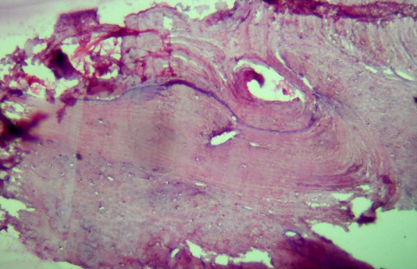
Temporomandibular joint (TMJ) forms a synovial articulation between the condyle and the cranium. It is a complex joint and shows hinge and gliding movements. Unlike other articulating heads, condyle grows with intramembranous ossification. TMJ is subjected to excessive loads throughout life as it supports essential functions such as mastication, deglutition, speech and respiration. Traumatic, neoplastic or non-neoplastic pathologies sometimes necessitate joint replacement therapy. Osteochondroma is one such benign tumour originating from condyle which requires surgical replacement of condyle with prosthesis. Various replacement methods have been designed in the past. Alloplastic grafts have been successfully used in joint replacement surgeries like hip joint, knee joint, etc. This case discussion supports the use of titanium-made condylar prosthesis for long-term functional stability of TMJ.
Figures


References
-
- Lichtenstein L. Bone tumors. 5th edn. St Louis, MO: CV Mosby, 1977
-
- Holmlund AB, Gynther GW, Reinholt FP. Surgical treatment of osteochondroma of the mandibular condyle in the adult. A 5-year follow-up. Int J Oral Maxillofac Surg 2004;33:549–53 - PubMed
-
- Dahlin DC, Unni KK. Bone tumours: general aspects and data on 8542 cases. Springfield, IL: Thomas, 1986:18
-
- Roychoudhury A, Bhatt K, Yadav R, et al. Review of osteochondroma of mandibular condyle and report of a case series. J Oral Maxillofac Surg 2011;69:2815–23 - PubMed
-
- Meng Q, Chen S, Long X, et al. The clinical and radiographic characteristics of condylar osteochondroma. Oral Surg Oral Med Oral Pathol Oral Radiol 2012;114:e66–74 - PubMed
Publication types
MeSH terms
LinkOut - more resources
Full Text Sources
Other Literature Sources
Medical
Miscellaneous