Review
. 2014 Apr;34(4):553-63.
doi: 10.1038/jcbfm.2014.18. Epub 2014 Feb 5.

Cerebral neovascularization in diabetes: implications for stroke recovery and beyond

Affiliations
Review

Cerebral neovascularization in diabetes: implications for stroke recovery and beyond

Adviye Ergul et al. J Cereb Blood Flow Metab. 2014 Apr.

Abstract

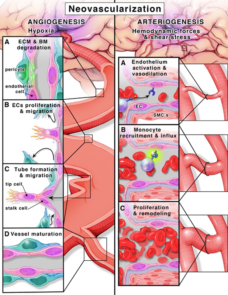
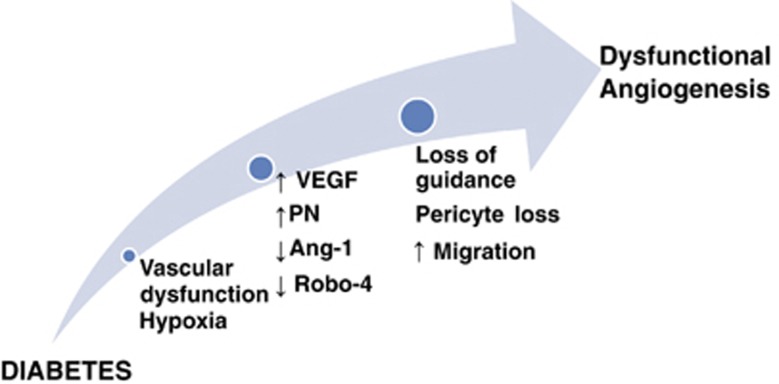
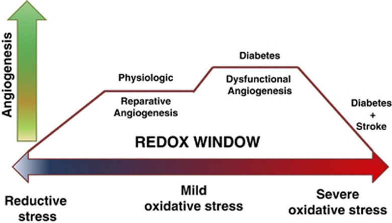
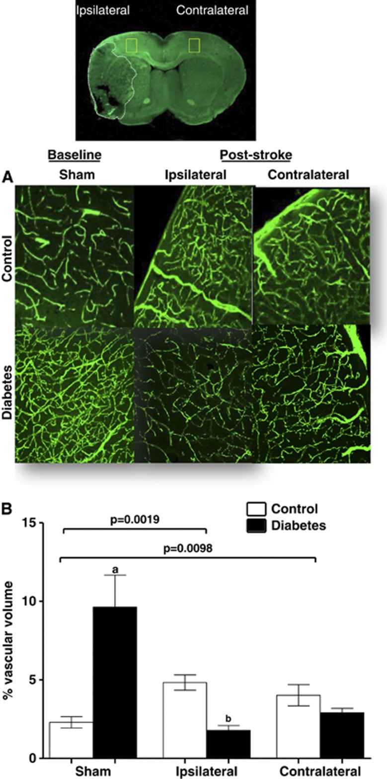
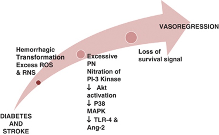
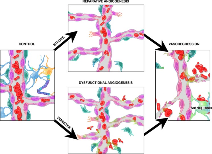
Neovascularization is an innate physiologic response by which tissues respond to various stimuli through collateral remodeling (arteriogenesis) and new vessel formation from existing vessels (angiogenesis) or from endothelial progenitor cells (vasculogenesis). Diabetes has a major impact on the neovascularization process but the response varies between different organ systems. While excessive angiogenesis complicates diabetic retinopathy, impaired neovascularization contributes to coronary and peripheral complications of diabetes. How diabetes influences cerebral neovascularization remained unresolved until recently. Diabetes is also a major risk factor for stroke and poor recovery after stroke. In this review, we discuss the impact of diabetes, stroke, and diabetic stroke on cerebral neovascularization, explore potential mechanisms involved in diabetes-mediated neovascularization as well as the effects of the diabetic milieu on poststroke neovascularization and recovery, and finally discuss the clinical implications of these effects.


Figures

Figure 1
Figure 1
Angiogenesis, formation of new vessels from existing ones (left panel). (A) ECM and BM degradation. Microenvironment hypoxia is a key stimulus for angiogenesis. Hypoxia activates the transcription factor HIF-1α that stimulates the transcription of pro-angiogenic molecules, growth factors (VEGF-A, VEGFR-2, FGF, and MMPs) and switches the environment balance toward angiogenic milieu (Ang-1/Ang-2). MMPs mediate ECM and BM degradation. (B) ECs proliferation and migration. Pericytes (blue) detach away from ECs (pink). The nearest EC to the highest gradient for VEGF transforms to tip cell that guide the following ECs (stalk cells) toward hypoxic tissue. (C) Tube formation and migration. Stalk cells proliferate and migrate forming a tube-like structure. (D) Vessel maturation. In the final stages, recruitment of pericytes promotes maturation and stabilization. Arteriogenesis, transformation of existing vessels into larger vessels in the normoxic tissue surrounding the ischemic area (right panel). (A) Endothelium activation and vasodilation. Hemodynamic forces and increased shear stress in collaterals activate vascular endothelium to proliferate and induce vasodilation. (B) Monocyte recruitment and influx. Upregulation of VCAM-1 and ICAM-1 and increased expression of MCP-1 and GM-CSF result in recruitment of monocytes. (C) Proliferation and remodeling. Monocytes transformed into macrophages secrete TNF-α and FGF that induce SMC (pink cells with pink nuclei) proliferation. Eventually, SMCs proliferation promotes outward remodeling and vessel maturation. Ang-1, angiopoietin-1; Ang-2, angiopoietin-2;, ECM, extracellular matrix; BM, basement membrane; ECs, endothelial cells; FGF, fibroblast growth factor; GM-CSF, granulocyte-macrophage colony-stimulating factor; HIF-1α, hypoxia inducible factor-1α; ICAM-1, intercellular adhesion molecule-1; MCP-1, monocyte chemotactic protein-1; MMPs, matrix metalloproteases; VEGF-A, vascular endothelial growth factor-A; VCAM-1, vascular cell adhesion molecule-1; SMCs, smooth muscle cells; TNF-α, tumor necrosis factor-α; VEGFR-2, VEGF receptor 2.
Figure 2
Figure 2
Cerebral angiogenesis in diabetes. A schematic illustrating the mechanisms by which diabetes induces cerebral dysfunctional angiogenesis. Diabetic hyperglycemia induces vascular dysfunction that creates a state of cerebral hypoxia. Diabetes-induced hypoxia triggers a series of events including (1) increased production of vascular endothelial growth factor (VEGF), (2) increased oxidative and nitrative stress (increased peroxynitrite (PN) formation), (3) decreased angiopoietin-1 (Ang-1), and (4) decreased expression of guidance molecule, Roundabout-4 (Robo-4). These events lead to pericyte loss, increase endothelial migration with loss of guidance that cumulatively increases dysfunctional cerebral angiogenesis.
Figure 3
Figure 3
Redox window and angiogenesis. A diagram representing the redox window concept: the tissue redox state ranging from reductive to oxidative levels is depicted on the X axis. Y axis is the angiogenesis process. Physiologic/reparative angiogenesis requires the tissue microenvironment to express mild levels of oxidative stress. Extreme levels of reductive or oxidative stress impair angiogenesis. Oxidative stress levels in diabetes depend on disease severity and promote dysfunctional angiogenesis. Adding stroke to diabetes greatly increases oxidative stress and corrupts the reparative angiogenic process after stroke.
Figure 4
Figure 4
Diabetes impairs poststroke neovascularization in the ipsilateral and contralateral hemispheres. Vascularization was assessed in the ipsilateral and contralateral hemispheres in control and diabetic animals 14 days after 90-minute occlusion of the middle cerebral artery. Sham animals were exposed to anesthesia and neck dissection was performed and sutured without occluding middle cerebral artery. Three-dimensional reconstruction of the fluorescein isothiocyanate (FITC)-stained vasculature was achieved analysis of the z-stack confocal images by the Volocity program. (A) Representative cortical images contrasting ipsilateral and contralateral zones in control and diabetic animals. (B) Plot depicting vascular volume across groups. aP<0.05 versus sham control or ipsilateral control and diabetes, bP<0.05 versus control. Data were analyzed with a 2 × 2 design for disease (control versus diabetes) and intervention (sham versus stroke) in the ipsilateral or contralateral hemispheres. There was a significant interaction indicating important differences in vascularization at baseline and after stroke in the diabetes group. n=6 to 9. (Modified from with permission from Lippincott, Williams, and Wilkins.)
Figure 5
Figure 5
Cerebral neovascularization in diabetes after stroke. A representative diagram illustrating the mechanisms by which vasoregression of cerebral vessels occurs in diabetes after stroke. When stroke is overlayed on diabetes pathology, there is greater hemorrhagic transformation and free iron accumulation that induces cell death. In addition, adding stroke injury to diabetes dramatically increases peroxynitrite (PN) formation and nitration of p85 regulatory subunit of PI3 kinase that downregulates the downstream pro-survival Akt pathway and activates the pro-apoptic p38MAP kinase pathway. Finally, increases in angiopoietin-2 (Ang-2) and activation of toll-like receptor 4 (TLR4) increase endothelial death and promote vasoregression in diabetes after stroke. ROS, reactive oxygen species; RNS, reactive nitrogen species.
Figure 6
Figure 6
Cerebral neovascularization in diabetes and stroke. Diabetes causes dysfunctional angiogenesis. Stroke stimulates reparative angiogenesis in the nondiabetic state. However, when stroke occurs in diabetes, survival signals are lost leading to vasoregression.

References

    1. Mink JW, Blumenschine RJ, Adams DB. Ratio of central nervous system to body metabolism in vertebrates: its constancy and functional basis. Am J Physiol. 1981;241:R203–R212. - PubMed
    1. Quaegebeur A, Lange C, Carmeliet P. The neurovascular link in health and disease: molecular mechanisms and therapeutic implications. Neuron. 2011;71:406–424. - PubMed
    1. Begley DJ, Brightman MW. Structural and functional aspects of the blood-brain barrier. Prog Drug Res. 2003;61:39–78. - PubMed
    1. Cipolla MJ.The cerebral circulationIn: Granger NGaJ, (ed). Integrated systems physiology: from molecule to function Morgan & Claypool Life Sciences: San Rafael (CA)2009 - PubMed
    1. Xu K, Lamanna JC. Chronic hypoxia and the cerebral circulation. J Appl Physiol. 2006;100:725–730. - PubMed

Publication types

MeSH terms

Substances

LinkOut - more resources