. 2014 Mar;45(3):857-64.
doi: 10.1161/STROKEAHA.113.004100. Epub 2014 Feb 4.

Essential role of program death 1-ligand 1 in regulatory T-cell-afforded protection against blood-brain barrier damage after stroke

Affiliations

Essential role of program death 1-ligand 1 in regulatory T-cell-afforded protection against blood-brain barrier damage after stroke

Peiying Li et al. Stroke. 2014 Mar.

Abstract

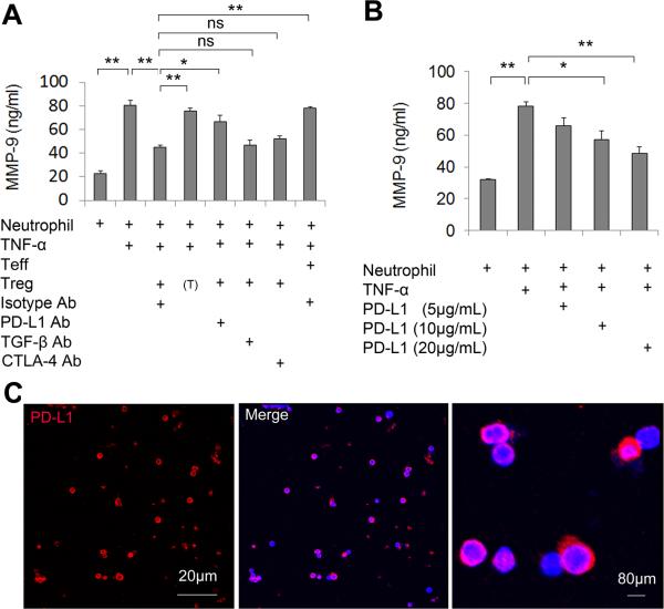
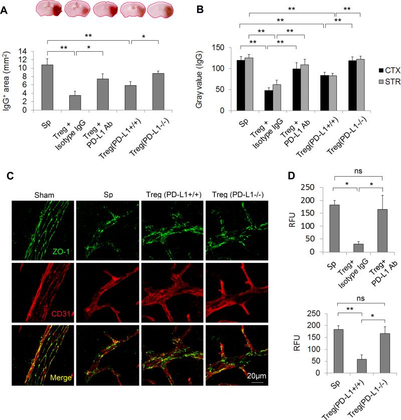
Background and purpose: Our recent research revealed that adoptively transferred regulatory T cells (Tregs) reduced acute ischemic brain injury by inhibiting neutrophil-derived matrix metalloproteinase-9 (MMP-9) and protecting against blood-brain barrier damage. The mechanisms underlying Treg interactions with neutrophils remain elusive. This study evaluates the contribution of program death 1-ligand 1 (PD-L1) to Treg-mediated neutrophil inhibition and neuroprotection after cerebral ischemia.

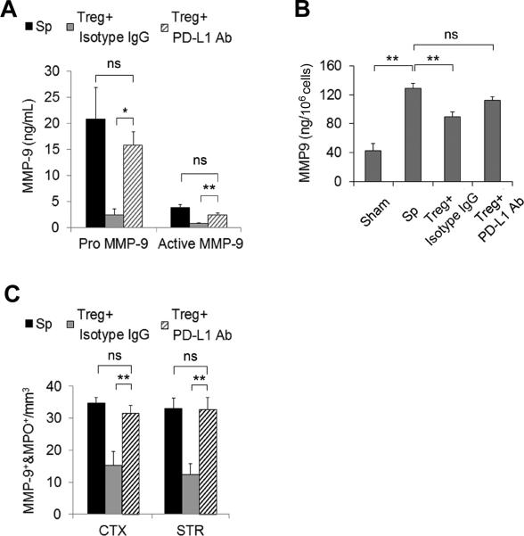
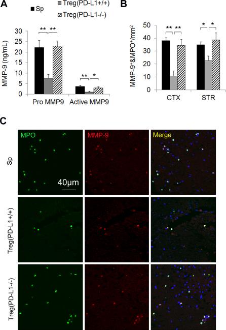
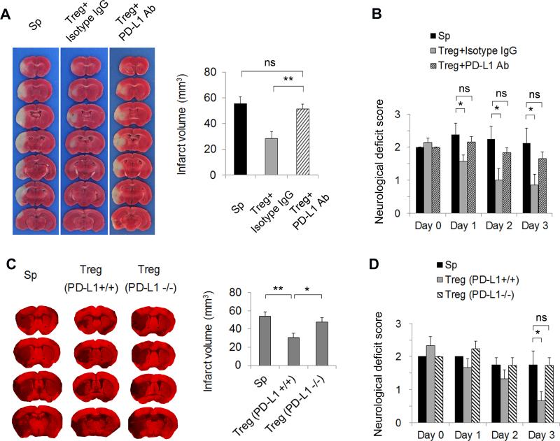
Methods: In vitro experiments were performed using a transwell system or a coculture system allowing cell-to-cell contact. Focal cerebral ischemia was induced in mice for 60 minutes. Tregs (2×10(6)) isolated from donor animals (wild-type or PD-L1-/-) were intravenously injected into ischemic recipients 2 hours after middle cerebral artery occlusion (MCAO). MMP-9 production, blood-brain barrier permeability, and brain infarct were assessed at 1 or 3 days after MCAO.

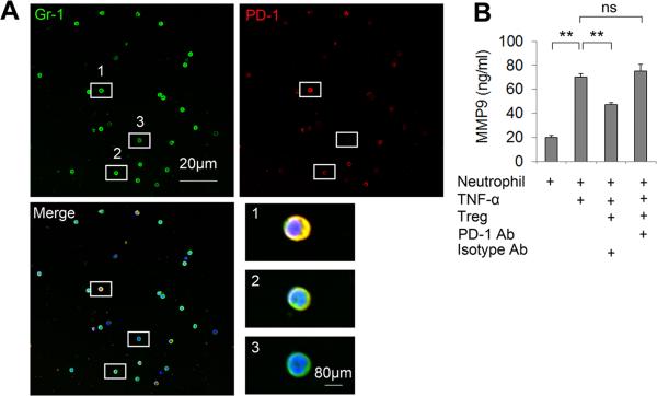
Results: In vitro experiments reveal that Treg-mediated inhibition of neutrophil MMP-9 required direct cell-to-cell contact. The suppression of MMP-9 was abolished when Tregs were pretreated with PD-L1 neutralizing antibodies or when neutrophils were pretreated with PD-1 antibodies. In vivo studies confirmed that intravenous administration of Tregs pretreated with PD-L1 antibodies or Tregs isolated from PD-L1-deficient mice failed to inhibit MMP-9 production by blood neutrophils 1 day after 60 minutes MCAO. Furthermore, the blood-brain barrier damage after MCAO was greatly ameliorated in PD-L1-competent Treg-treated mice but not in PD-L1-compromised Treg-treated mice. Consequently, PD-L1 dysfunction abolished Treg-mediated brain protection and neurological improvements 3 days after MCAO.

Conclusions: PD-L1 plays an essential role in the neuroprotection afforded by Tregs against cerebral ischemia by mediating the suppressive effect of Tregs on neutrophil-derived MMP-9.

Keywords: matrix metalloproteinase 9; neutrophils; programmed cell death 1 ligand 1; regulatory T-cells; stroke.


Figures

Figure 1
Figure 1. Tregs inhibit neutrophil-derived MMP-9 through PD-L1 in in vitro cultures
(A) Isolated Tregs were stimulated with anti-CD3Ab and anti-CD28 Abs for 3 days and pretreated with various neutralizing Abs (anti-PD-L1, -TGF-β or -CTLA-4; 20 μg/mL) or isotype control IgG (20 μg/mL) for 1 h. Stimulated Tregs were co-cultured with neutrophils and then exposed to TNF-α (100 ng/mL) for 24 h. MMP-9 in the culture medium was measured by ELISA. (T) indicates that neutrophils were cultured in transwells. (B) Neutrophils were incubated with PD-L1 protein at indicated concentrations. Cells were then exposed to TNF-α (100 ng/mL) for 24 h. MMP-9 in the culture medium was measured by ELISA. n=6/group in 3 independent experiments. *P<0.05, **P<0.01. (C) Immunocytochemical staining of PD-L1 on Tregs. Red: PD-L1. Blue: DAPI.
Figure 2
Figure 2. PD-1 expressed on neutrophils mediates Treg-neutrophil interactions
(A) Double staining of Gr-1 (a neutrophil marker) and PD-1 on neutrophils extracted from mice at 3 days after MCAO. High magnification images showing neutrophils with varying levels of PD-1 expression. 1, high expression; 2, low expression; 3, no expression. (B) Neutrophils isolated from mouse blood and bone marrow were pre-incubated with 20 μg/mL PD-1 Abs or 20 μg/mL isotype control IgG and then co-cultured with Tregs. Cells were then exposed to TNF-α (100 ng/mL) for 24 h. MMP-9 in the culture medium was measured by ELISA. n=6/group. *P<0.05, **P<0.01, ns: not significant.
Figure 3
Figure 3. PD-L1 blocking Abs abolish Treg-mediated inhibition of the rise in MMP-9 after MCAO
Tregs (2×10⁶/animal) were pre-incubated with PD-L1 neutralization Abs or isotype control IgG at 20 μg/mL and then adoptively transferred into MCAO animals after 2 h of reperfusion. (A) Quantification of plasma MMP-9 at 24 h after MCAO (n=7/group). (B) Quantification of blood neutrophil-derived MMP-9 at 24 h after MCAO (n=4/group). Neutrophils were isolated from the blood of MCAO and sham mice at 24 h after operation. MMP-9 was measured in the neutrophil lysate by ELISA. (C) The infiltration of MMP-9+MPO+ neutrophils into the cortex (CTX) and striatum (STR) was quantified at 3 days after MCAO (n=5/group). *P<0.05, **P<0.01, ns: not significant. Sp = splenocytes
Figure 4
Figure 4. Tregs prepared from PD-L1 deficient mice fail to inhibit the rise of blood MMP-9 after MCAO
Tregs were prepared from PD-L1 knockout mice and then adoptively transferred into MCAO animals after 2 h of reperfusion. (A) Quantification of plasma MMP-9 1 day after MCAO (n=5/group). (B) The infiltration of MMP-9+MPO+ neutrophils into the cortex (CTX) and striatum (STR) was quantified at 3 days after MCAO (n=5/group). (C) Representative images of MMP-9 and MPO double staining on brain sections. Images are representative of brain sections from five mice in each group. Scale bar represents 40 μm *P<0.05, **P<0.01. Sp = splenocytes.
Figure 5
Figure 5. PD-L1 is critical for Treg-afforded BBB preservation after MCAO
To block PD-L1 signaling in Tregs, Tregs were pre-incubated with PD-L1 neutralizing Abs or prepared from PD-L1 knockout mice and then adoptively transferred into MCAO animals after 2 h of reperfusion. Isotype IgG-treated or wild-type Tregs were used as controls, respectively. (A-B) Quantification of IgG extravasation at 24 h after MCAO. (A) Quantification of endogenous IgG positive area determined by immunohistochemical staining of mouse IgG. n=5/group. (B) Quantification of gray values of IgG immunostaining in the cortex (CTX) and striatum (STR). n=5/group. (C) Representative Z-stack confocal images of the tight junction protein ZO-1 and endothelial cell marker CD31 in brain sections obtained 1 day after MCAO. Images are representative of brain sections from four mice in each group. (D) Cadaverine-Alexa-555 (950Da) was injected intravenously 22 h post-MCAO. Fluorescence intensities in brain lysates from the infarct area were measured after 2 h of circulation. n=5/group. *P<0.05, **P<0.01. Sp = splenocytes.
Figure 6
Figure 6. PD-L1 is critical for Treg-afforded neuroprotection after MCAO
(A-B) Tregs were pre-incubated with PD-L1 neutralizing Abs or isotype control IgG at 20 μg/mL and then adoptively transferred into MCAO animals after 2 h of reperfusion. (A) Representative TTC staining of brain infarct and quantification of infarct volume at 3 days after MCAO. n=7/group. (B) Neurological deficit scores over 3 days after MCAO. n=7/group. (C-D) Tregs were prepared from PD-L1 knockout mice and then adoptively transferred into MCAO animals after 2 h of reperfusion. (C) Representative MAP2 staining of brain infarct and quantification of infarct volume at 3 days after MCAO. n=5/group. (D) Neurological deficits scores over 3 days after MCAO. n=5/group. *P<0.05, **P<0.01, ns: not significant. Sp = splenocytes.

References

    1. Fernandez-Lopez D, Faustino J, Daneman R, Zhou L, Lee SY, Derugin N, et al. Blood-brain barrier permeability is increased after acute adult stroke but not neonatal stroke in the rat. J Neurosci. 2012;32:9588–9600. - PMC - PubMed
    1. Asahi M, Wang X, Mori T, Sumii T, Jung JC, Moskowitz MA, et al. Effects of matrix metalloproteinase-9 gene knock-out on the proteolysis of blood-brain barrier and white matter components after cerebral ischemia. J Neurosci. 2001;21:7724–7732. - PMC - PubMed
    1. Rosell A, Ortega-Aznar A, Alvarez-Sabin J, Fernandez-Cadenas I, Ribo M, Molina CA, et al. Increased brain expression of matrix metalloproteinase-9 after ischemic and hemorrhagic human stroke. Stroke; a journal of cerebral circulation. 2006;37:1399–1406. - PubMed
    1. Park KP, Rosell A, Foerch C, Xing C, Kim WJ, Lee S, et al. Plasma and brain matrix metalloproteinase-9 after acute focal cerebral ischemia in rats. Stroke. 2009;40:2836–2842. - PMC - PubMed
    1. Rosell A, Cuadrado E, Ortega-Aznar A, Hernandez-Guillamon M, Lo EH, Montaner J. Mmp-9-positive neutrophil infiltration is associated to blood-brain barrier breakdown and basal lamina type iv collagen degradation during hemorrhagic transformation after human ischemic stroke. Stroke. 2008;39:1121–1126. - PubMed

Publication types

MeSH terms