. 2014 Feb 4;3(2):e146.
doi: 10.1038/mtna.2013.75.

In vitro Inactivation of Latent HSV by Targeted Mutagenesis Using an HSV-specific Homing Endonuclease

Affiliations

In vitro Inactivation of Latent HSV by Targeted Mutagenesis Using an HSV-specific Homing Endonuclease

Martine Aubert et al. Mol Ther Nucleic Acids. .

Abstract

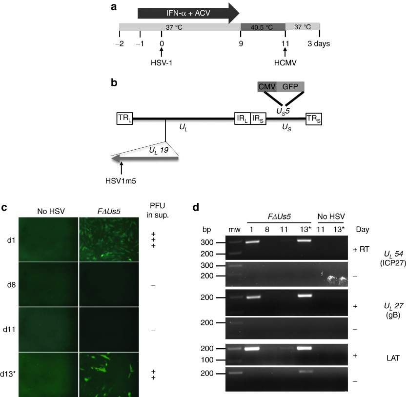
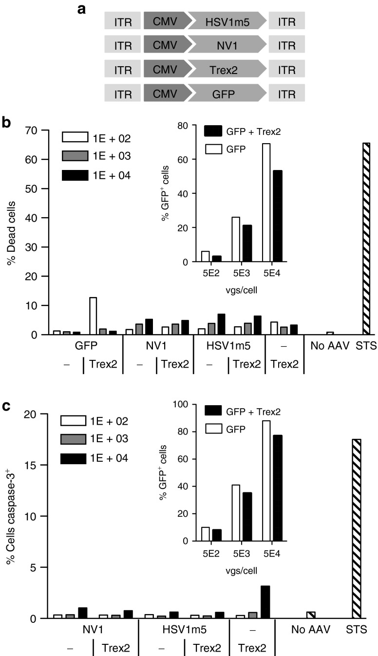
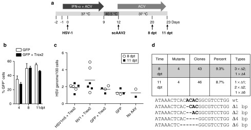
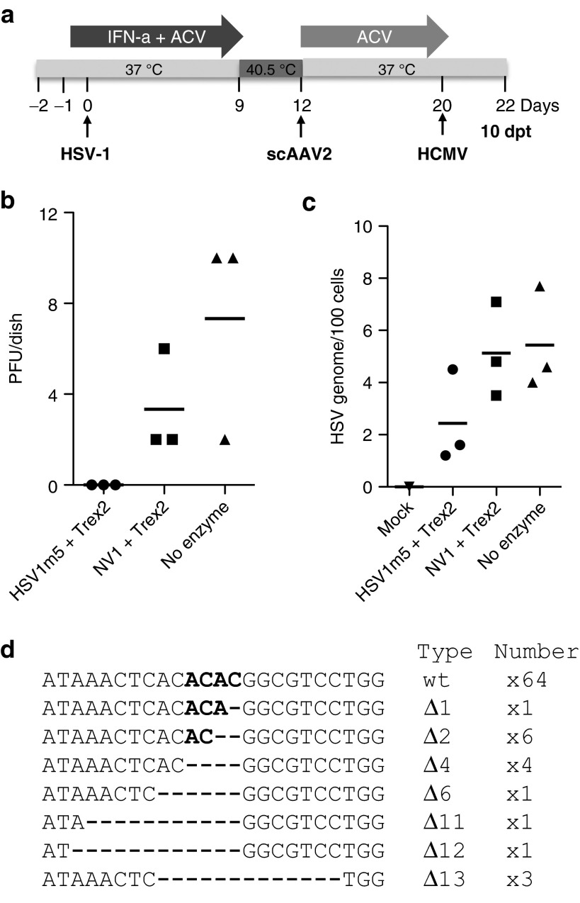
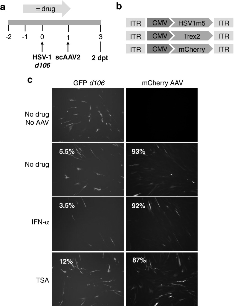
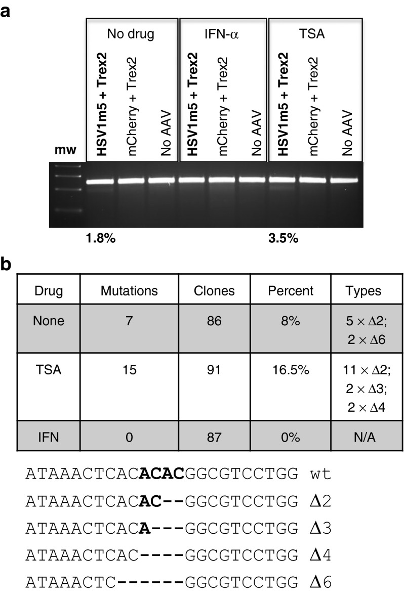
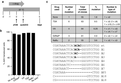
Following acute infection, herpes simplex virus (HSV) establishes latency in sensory neurons, from which it can reactivate and cause recurrent disease. Available antiviral therapies do not affect latent viral genomes; therefore, they do not prevent reactivation following therapy cessation. One possible curative approach involves the introduction of DNA double strand breaks in latent HSV genomes by rare-cutting endonucleases, leading to mutagenesis of essential viral genes. We tested this approach in an in vitro HSV latency model using the engineered homing endonuclease (HE) HSV1m5, which recognizes a sequence in the HSV-1 gene UL19, encoding the virion protein VP5. Coexpression of the 3'-exonuclease Trex2 with HEs increased HE-mediated mutagenesis frequencies up to sixfold. Following HSV1m5/Trex2 delivery with adeno-associated viral (AAV) vectors, the target site was mutated in latent HSV genomes with no detectable cell toxicity. Importantly, HSV production by latently infected cells after reactivation was decreased after HSV1m5/Trex2 exposure. Exposure to histone deacetylase inhibitors prior to HSV1m5/Trex2 treatment increased mutagenesis frequencies of latent HSV genomes another two- to fivefold, suggesting that chromatin modification may be a useful adjunct to gene-targeting approaches. These results support the continuing development of HEs and other nucleases (ZFNs, TALENs, CRISPRs) for cure of chronic viral infections.Molecular Therapy-Nucleic Acids (2014) 3, e1; doi:10.1038/mtna.2013.75; published online 4 February 2014.


Figures

Figure 1
Figure 1
In vitro model of herpes simplex virus (HSV) latency. (a) Timeline of the establishment of HSV latency in human fibroblasts (HF) and human cytomegalovirus (HCMV) reactivation of latent HSV. ACV, acyclovir; IFN, interferon. (b) Schematic representation of the HSV-1 FΔUs5 genome. The long terminal and internal repeats (TRL and IRL), and the internal and terminal short repeats (TRS and IRS) bordering the unique long (UL) and unique short (US) regions are shown. A green flourescent protein (GFP) expression cassette is inserted in the Us5 gene. The location of the target sequence recognized by the HSV-specific HE (HSV1m5) in the UL19 gene is indicated. (c) GFP expression as assessed by fluorescence microscopy at 1, 8, 11, and 13 days postinfection with FΔUs5. At day 11, cells were infected with HCMV AD169 (multiplicity of infection (MOI) = 1) to reactivate latent HSV (×10 magnification). *Indicates sample analyzed after HSV reactivation. Presence of progeny virus in the cell culture supernatant is indicated on the right (PFU: Plaque forming unit; +++ indicates ≥ 100 PFU/ml; ++ indicates ≥ 10 PFU/ml; - indicates no virus detected). (d) Reverse transcription polymerase chain reaction (RT-PCR) products for the immediate early gene UL54 encoding ICP27, late gene UL27 encoding glycoprotein B (gB) and latency-associated transcript (LAT) were generated from total RNA extracted at 1, 8, 11, 13 days postinfection with FΔUs5 and resolved on a 2% agarose gel. Reverse transcriptase (RT) was either added (+) or omitted (−). bp, base pair; mw, molecular weight size marker. *Indicates sample analyzed after HSV reactivation.
Figure 2
Figure 2
Lack of homing endonuclease (HE)-induced toxicity. (a) Schematic representation of scAAV vectors constructs for transgene delivery. CMV, cytomegalovirus early promoter; ITR, inverted terminal repeat. Human fibroblasts (HF) transduced for 4 days with scAAV2 delivery vectors expressing the indicated protein (GFP, NV1, or HSV1m5 in the presence or absence of Trex2, or Trex2 alone) at a multiplicity of infection (MOI) of 1 × 102, 1 × 103, or 1 × 104 vgs/cells per vector or untransduced (no AAV) were subjected to flow cytometry analysis following staining with either (b) live/dead stain or (c) anti-activated caspase-3 antibody. Untransduced control cells were treated with 0.5 µmol/l staurosporine (STS) 24 hours prior to analysis to provide a positive control for death and apoptosis. The insert graph shows the levels of transduction in control cells transduced with either scAAV2-GFP only or scAAV2-GFP and scAAV2-Trex2 at the indicated MOI. GFP fluorescence in control cells was measured by flow cytometry.
Figure 3
Figure 3
Targeted mutagenesis of latent herpes simplex virus (HSV) genomes. (a) Timeline of HSV latency establishment in human fibroblasts (HF) and exposure to scAAV2 delivery vectors. HF were plated (105/well), pretreated with ACV (30 µmol/l) and IFN-α (200 U/ml), infected with HSV-1 FΔUs5 at a MOI of 2.5 and maintained in the presence of drugs for 9 days with daily replenishment. The drugs were removed and the incubation temperature increased to 40.5 °C to maintain HSV latency. HSV-infected HFs were then transduced with scAAV2 expressing Trex2 and either HSV-1m2, NV1, or GFP at a MOI of 104 genomes/cell each and maintained in culture medium with ACV for either 8 or 11 additional days. ACV, acyclovir; IFN, interferon. (b) Assessment of transduction levels with scAAV vectors by flow cytometry for GFP at 4, 8, and 11 days posttransduction in duplicate dishes of HSV-latently infected HF exposed to scAAV2-GFP alone or in combination with scAAV2-Trex2. Mean + SD. (c) Quantitation of HSV genomes in latently-infected HF exposed to scAAV2 vectors by ddPCR in triplicate dishes at 8 (open circle) or 11 (filled circle) days posttransduction. The bars represent the mean of the combined six dishes for each condition. (d) At 8 and 11 days posttransduction, the presence of mutations at the target site of HSV1m5 in HSV genomes from HSV1m5-/Trex2-treated cells was analyzed by polymerase chain reaction (PCR) amplicon sequencing. The HSV region containing the target site was PCR amplified from total genomic DNA obtained from triplicate dishes of HSV-latently infected HF treated with HSV1m5 and Trex2. The PCR amplicons were pooled, cloned and sequenced from individual bacterial colonies. The percent and type of mutations found are presented. Wt, wild-type sequence of the HSV1m5 target site. Bold indicates the four nucleotides constituting the 3′ overhangs generated by the DNA DSB; - indicates a deleted nucleotide.
Figure 4
Figure 4
Impact of homing endonuclease (HE)-mediated mutagenesis on herpes simplex virus (HSV) reactivation. (a) Timeline of HSV latency establishment in human fibroblasts (HF), exposure to scAAV2 delivery vectors and reactivation following CMV infection. Triplicate dishes of latently-infected HF transduced at a multiplicity of infection (MOI) of 104 genomes/cell/vector with scAAV2 delivery vectors expressing the indicated transgene were reactivated at day 20 (8 dpt) by infection with HCMV AD169 at a MOI of 1 for 2 days in the absence of ACV. At 2 days postinfection with human cytomegalovirus (HCMV) (day 22; 10 dpt). ACV, acyclovir; IFN, interferon. (b) The presence of virion progeny in culture supernatants was assessed by plaque assay titration and (c) the number of intracellular HSV genomes was measured by ddPCR. Bars indicate the mean from the triplicate samples. (d) The HSV region containing the target site was PCR amplified from total genomic DNA obtained from triplicate dishes of reactivated HSV infected HF treated with HSV1m5 and Trex2. The PCR amplicons were pooled, cloned, and sequenced from individual bacterial colonies. The nature and number of the mutations found are indicated on the right side of each sequence. Δ: deletion; wt, wild-type sequence of the HSV1m5 target site. Bold indicates the four nucleotides constituting the 3′ overhang generated by the DNA DSB; - indicates a deleted nucleotide.
Figure 5
Figure 5
Impact of epigenetic modification of the herpes simplex virus (HSV) genome on homing endonuclease (HE)-mediated mutagenesis. (a) Schematic of experimental timeline. MRC5 cells left untreated or drug-treated with IFN-α (200 U/ml) or trichostatin-A (TSA; 100 nmol/l), infected with recombinant HSV-1 d106 at a multiplicity of infection (MOI) of 0.5 were transduced with scAAV2-HSV1m5 and scAAV2-Trex2 or scAAV2-mCherry and scAAV2-Trex2 at a MOI of 5 × 104 genomes/cell/vector for 2 days in the absence of drug. (b) Schematic representation of the scAAV delivery vectors used. CMV, cytomegalovirus early promoter; ITR, inverted terminal repeat. (c) Fluorescence microscopy analysis of GFP (HSV d106 gene expression; left column) and mCherry (AAV transduction; right column) in control d106-infected MRC5 at a MOI of 5 × 104 genomes/cell/vector for 2 days. Percent of cells expressing GFP and mCherry fluorescence was also quantified by flow cytometry (percent of positive cells indicated in the upper left corner of each panel).
Figure 6
Figure 6
Mutational events in herpes simplex virus (HSV)-infected human fibroblasts (HF) treated with trichostatin-A (TSA) or interferon (IFN)-α. (a) Polymerase chain reaction (PCR) amplicons of the HSV region containing the HSV1m5 target site were treated with Surveyor nuclease, and digested products were separated on a 3% agarose gel and quantified using ImageJ. mw: molecular weight size ladder. The percent of sequence modification detected by this assay is indicated. (b) HSV1m5 target sequence analysis. The HSV region containing the HSV1m5 target site was PCR amplified from total genomic DNA obtained at 2 days post-AAV transduction. PCR amplicons were cloned and sequenced from individual bacterial colonies. The frequency and nature of the mutations found are presented. N/A, not applicable, Δ: deletion; wt, wild-type target sequence. Bold indicates the four nucleotides constituting the 3′ overhang generated by the DNA DSB.
Figure 7
Figure 7
Targeted mutagenesis in herpes simplex virus (HSV)-infected cells treated with histone deacetylase inhibitor (HDACi). (a) Schematic of experimental timeline. MRC5 cells were infected with recombinant HSV-1 d106 at a multiplicity of infection (MOI) of 0.5 in the absence or presence of drug, transduced with scAAV2 at a MOI of 5 × 104 genomes/cell/vector in the absence of drug for 4 days prior to analysis for mutational events. (b) Flow cytometry for green flourescent protein (GFP) fluorescence in control uninfected MRC5 cells cultured in the absence (no drug) or presence of the indicated HDACi for 2 days, then either left untransduced (no AAV) or transduced with scAAV2-GFP and scAAV2-Trex2 at a MOI of 5 × 104 genomes/cell/vector for 4 days in the absence of drug. (c) HSV1m5 target sequence analysis. The HSV region containing the HSV1m5 target site was PCR amplified using total genomic DNA obtained at 4 days post-AAV transduction from d106-infected MRC5 exposed to HSV1m5 and Trex2 in the absence (none) or presence of the indicated HDACi. PCR amplicons were cloned and sequenced from individual bacterial colonies. The percent and nature of mutations in the HSV1m5 target site are shown. wt, wild-type sequence of the HSV1m5 target site. Bold indicates the four nucleotides constituting the 3′ overhang generated by the DNA DSB. Δ: deletion. HDACi used: trichostatin A (TSA 100 nmol/l), valproic acid (VA 1 mmol/l), 3-deazaneplanocin (DZNep 5 µmol/l), sodium butyrate (NaBu 5 mmol/l).

References

    1. Schiffer JT, Corey L. New concepts in understanding genital herpes. Curr Infect Dis Rep. 2009;11:457–464. - PMC - PubMed
    1. Xu F, Sternberg MR, Kottiri BJ, McQuillan GM, Lee FK, Nahmias AJ, et al. Trends in herpes simplex virus type 1 and type 2 seroprevalence in the United States. JAMA. 2006;296:964–973. - PubMed
    1. Berrington WR, Jerome KR, Cook L, Wald A, Corey L, Casper C. Clinical correlates of herpes simplex virus viremia among hospitalized adults. Clin Infect Dis. 2009;49:1295–1301. - PMC - PubMed
    1. Brown EL, Gardella C, Malm G, Prober CG, Forsgren M, Krantz EM, et al. Effect of maternal herpes simplex virus (HSV) serostatus and HSV type on risk of neonatal herpes. Acta Obstet Gynecol Scand. 2007;86:523–529. - PubMed
    1. Serwadda D, Gray RH, Sewankambo NK, Wabwire-Mangen F, Chen MZ, Quinn TC, et al. Human immunodeficiency virus acquisition associated with genital ulcer disease and herpes simplex virus type 2 infection: a nested case-control study in Rakai, Uganda. J Infect Dis. 2003;188:1492–1497. - PubMed