. 2013 May 1;51(9):1899-1911.
doi: 10.1002/pola.26574.

Modeling Controlled Photodegradation in Optically Thick Hydrogels

Affiliations

Modeling Controlled Photodegradation in Optically Thick Hydrogels

Mark W Tibbitt et al. J Polym Sci A Polym Chem. .

Abstract

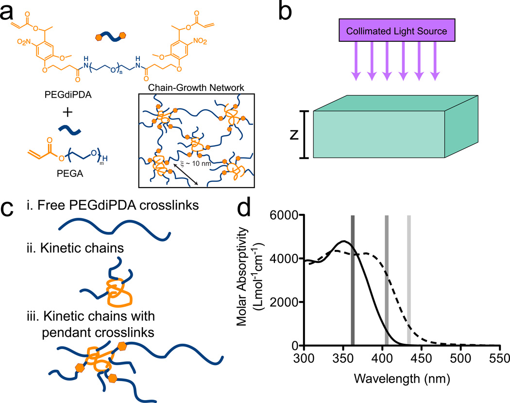
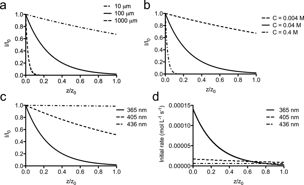
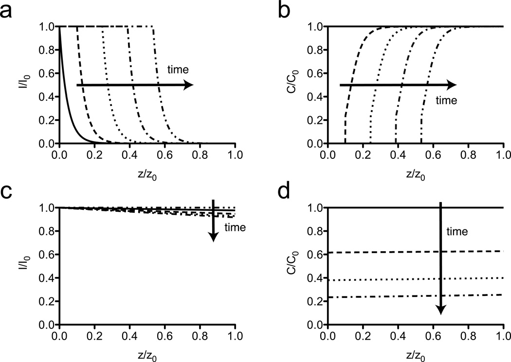
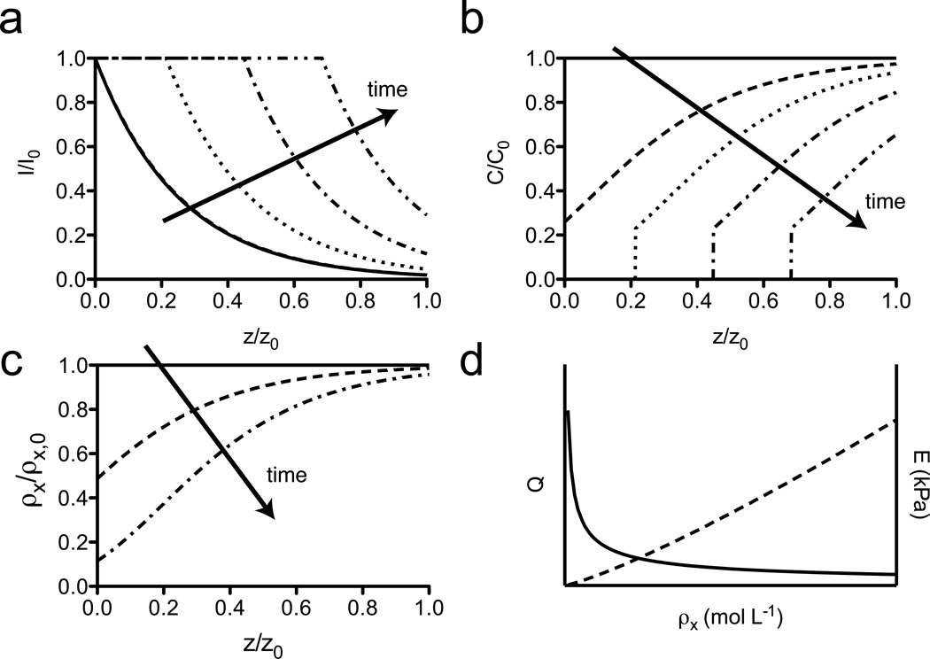
There is a growing interest in developing dynamically responsive hydrogels whose material properties are modulated by environmental cues, including with light. These photoresponsive hydrogels afford spatiotemporal control of material properties through an array of photoaddition and photodegradation reactions. For photoresponsive hydrogels to be utilized most effectively in a broad range of applications, the photoreaction behavior should be well understood, enabling the design of dynamic materials with uniform or anisostropic material properties. Here, a general statistical-kinetic model has been developed to describe controlled photodegradation in hydrogel polymer networks containing photolabile crosslinks. The heterogeneous reaction rates that necessarily accompany photochemical reactions were described by solving a system of partial differential equations that quantify the photoreaction kinetics in the material. The kinetics were coupled with statistical descriptions of network structure in chain polymerized hydrogels to model material property changes and mass loss that occur during the photodegradation process. Finally, the physical relevance of the model was demonstrated by comparing model predictions with experimental data of mass loss and material property changes in photodegradable, PEG-based hydrogels.

Keywords: degradable materials; hydrogels; modeling; photodegradation; photoresponsive materials.


Figures

Figure 1
Figure 1. Photodegradable hydrogel system
(a) Photodegradable PEG based hydrogels were formed through redox-initiated free radical polymerization of the photodegradable crosslinker (PEGdiPDA) with a co-monomer (PEGA). (b) Upon exposure to collimated irradiation, these gels degrade as the photolabile o-nitrobenzyl ether (NBE) cleaves leading to mass loss and, ultimately, erosion. (c) Mass loss results from the release of cleavage products from the network that predominantly fall into three categories: i. free PEGdiPDA crosslinks, ii. kinetic chains, and iii. kinetic chains with pendant crosslinks. Once a sufficient fraction of NBE moieties are cleaved in a local region, the gel undergoes reverse gelation where the polymers no longer form an infinite molecular weight network and the local material erodes from the gel. (d) Photodegradation can be achieved at wavelengths where NBE absorbs light (solid line). In this work, 365 nm, 405 nm, and 436 nm were employed for degradation. The mechanism of mass loss of the gels was dictated by the total absorptivity of the degradable hydrogels, which depends on the molar absorptivity of NBE (solid line), the molar absorptivity of the cleavage product (NBP; dashed line), the concentrations of photoactive species, the thickness of the gel, and the wavelength of irradiation. Molar absorptivity data previously reported in ref. .
Figure 2
Figure 2. Model predictions of light intensity profiles and kinetic rates
The incident irradiation is attenuated in the hydrogel on account of the absorbing NBE within the PEGdiPDA hydrogels. (a) Increased thickness of the hydrogel ([NBE] = 0.04 M; λ = 365 nm; I0 = 10 mW cm−2) leads to decreased penetration depth of the light through the gel (z = 10 µm, dash-dot line; z = 100 µm, solid line; z = 1000 µm, dashed line). (b) Similarly, increased concentration of the NBE moieties in the gel (z = 100 µm; λ = 365 nm; I0 = 10 mW cm−2) leads to decreased penetration depth of the light through the gel ([NBE] = 0.004M, dashed line; [NBE] = 0.04M, solid line; [NBE] = 0.4M, dash-dot line). (c) The penetration depth of the light in the gel is also dependent on the wavelength of incident irradiation ([NBE] = 0.04 M; z = 100 µm; I0 = 10 mW cm−2). Here, 365 nm irradiation (solid line) is strongly attenuated owing to the high molar absorptivity of NBE at 365 nm (ɛNBE = 4300 L mol−1 cm−1). 405 nm irradiation (dashed line) leads to increased penetration depth (ɛNBE = 720 L mol−1 cm−1) while 436 nm irradiation (dash-dot line) is nearly uniform through the depth of the gel (ɛNBE = 16 L mol−1 cm−1). (d) The initial photocleavage rate as a function of depth is dependent on the local light intensity and the wavelength of irradiation, through changes in absorbance and quantum yield. ([NBE] = 0.04 M; I0 = 10 mW cm−2; z = 100 µm; λ = 365 nm, solid line; λ = 405 nm, dashed line; λ = 436 nm, dash-dot line)
Figure 3
Figure 3. Model predictions of surface and bulk erosion of photodegradable hydrogels
Surface erosion and bulk degradation are both observed in silico for photodegradable hydrogels depending on the total absorbance of the material. (a) PEGdiPDA hydrogels with high absorbance (A = 10; [NBE] = 0.23 M; z = 100 µm) severely attenuate the incident irradiation (λ = 365 nm; I0 = 10 mW cm−2), which leads to a front of light moving through the depth of the material as degradation proceeds. (arrows indicate the direction of increasing time; t = 0 min, solid line; t = 22.5 min, dashed line; t = 45 min, dotted line; t = 67.5 min, dash-dot line; t = 90 min, dash-double dot line) (b) Photodegradation erodes the surface of the material, where light remains at a high intensity. Erosion is indicated by C/C0 = 0. In this surface erosion case, the strongly coupled equations (Eqs. 1 – 3) lead to a propagation of light and mass loss through the depth of the gel with time. (t = 0 min, solid line; t = 22.5 min, dashed line; t = 45 min, dotted line; t = 67.5 min, dash-dot line; t = 90 min, dash-double dot line) (c) PEGdiPDA hydrogels with low absorbance (A = 0.01; [NBE] = 0.0056 M; z = 25 µm) allow incident irradiation (λ = 405 nm; I0 = 25 mW cm−2) to penetrate through the full depth of the gel uniformly, which results in bulk degradation of the material. (t = 0 min, solid line; t = 7.5 min, dashed line; t = 15 min, dotted line; t = 22.5 min, dash-dot line; t = 30 min, dash-double dot line) (d) As light penetrates the material uniformly, the reaction rate is uniform through the depth of the gel, which results in an even decrease in [NBE] through the depth of the gel, which is indicative of bulk degradation. At late time points, the whole material erodes. (t = 0 min, solid line; t = 7.5 min, dashed line; t = 15 min, dotted line; t = 22.5 min, dash-dot line; t = 30 min, dash-double dot line) For all model calculations N = 20.
Figure 4
Figure 4. Model predictions of photodegradation in PEGdiPDA hydrogels
For PEGdiPDA hydrogels with intermediate absorbance (A ~ 0.1 – 10), degradation is observed to be a mixture of surface erosion and bulk degradation. (a) In these materials, light is mostly confined to the surface volume of the gel, while a portion of the light is able to penetrate the full thickness, leading to non-neglibile cleavage of NBE moieties. (b) As time progresses the light front moves through the gel surface, eroding the material while anisotropically patterning the full depth of the gel. (arrows indicate the direction of increasing time; t = 0 min, solid line; t = 7.5 min, dashed line; t = 15 min, dotted line; t = 22.5 min, dash-dot line; t = 30 min, dash-double dot line) (c) The anisotropic patterning caused by this phenomenon is predicted to generate materials with gradients in crosslinking density as a function of depth. (t = 0 min, solid line; t = 2 min, dashed line; t = 6 min, dashed-dot line) (d) Since the volumetric swelling ratio, Q, and Young’s modulus, E, of PEG hydrogels depend on the crosslinking density, ρx, anisotropies in crosslinking density are readily generated to create spatially varying materials, that can be difficult to synthesize through traditional fabrication routes. For all model calculations, N = 20; [NBE] = 0.04 M; z = 100 µm; λ = 365 nm; I0 = 10 mW cm−2; A = 1.7.
Figure 5
Figure 5. Model predictions compared to experimental degradation of PEGdiPDA hydrogels
The photodegradation model was employed to predict mass loss and material property changes in chain polymerized, PEGdiPDA hydrogels. (a) Fractional mass loss for two formulations ([NBE] = 0.04 M; z = 1500 µm; triangles – experimental, previously reported data found in ref. ; dashed line – prediction and [NBE] = 0.058 M; z = 1500 µm; squares – experimental, previously reported data found in ref. ; solid line – prediction) in response to irradiation (λ = 365 nm; I0 = 20 mW/cm2) was predicted by the statistical-kinetic mass loss model. (b) Fractional mass loss for PEGdiPDA hydrogels ([NBE] = 0.04 M) in response to irradiation (λ = 405 nm at I0 = 25 mW/cm2) also compared well with predictions. (triangles – experimental; solid line – prediction) (c) The depth of channels patterned into the surface of PEGdiPDA hydrogels ([NBE] = 0.04 M; λ = 365 nm; I0 = 10 mW/cm2) was predicted, except at short irradiation times. (triangles – experimental, previously reported data found in ref. ; solid line – prediction) (d) A gradient in surface elasticity generated by photomasking was also predicted by the statistical-kinetic model. (triangles – experimental, previously reported data found in ref. ; solid line – prediction, line connects discrete model predictions) For all model calculations, N = 20.

Similar articles

Cited by

References

    1. Peppas N, Hilt J, Khademhosseini A, Langer R. Hydrogels in biology and medicine: From molecular principles to bionanotechnology. Adv Mater. 2006;18:1345–1360.
    1. Liechty WB, Kryscio DR, Slaughter BV, Peppas NA. Polymers for drug delivery systems. Annu Rev Chem Biomol Eng. 2010;1:149–173. - PMC - PubMed
    1. Tibbitt MW, Anseth KS. Hydrogels as Extracellular Matrix Mimics for 3D Cell Culture. Biotechnol Bioeng. 2009;103:655–663. - PMC - PubMed
    1. Lutolf MP, Hubbell JA. Synthetic biomaterials as instructive extracellular microenvironments for morphogenesis in tissue engineering. Nat Biotechnol. 2005;23:47–55. - PubMed
    1. Hilt JZ, Gupta AK, Bashir R, Peppas NA. Ultrasensitive Biomems Sensors Based on Microcantilevers Patterned with Environmentally Responsive Hydrogels. Biomedical Microdevices. 2003;5:177–184.