. 2014 Feb;141(4):878-88.
doi: 10.1242/dev.101709.

Machine learning classification of cell-specific cardiac enhancers uncovers developmental subnetworks regulating progenitor cell division and cell fate specification

Affiliations

Machine learning classification of cell-specific cardiac enhancers uncovers developmental subnetworks regulating progenitor cell division and cell fate specification

Shaad M Ahmad et al. Development. 2014 Feb.

Abstract

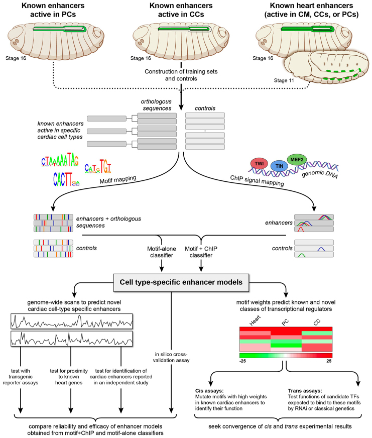
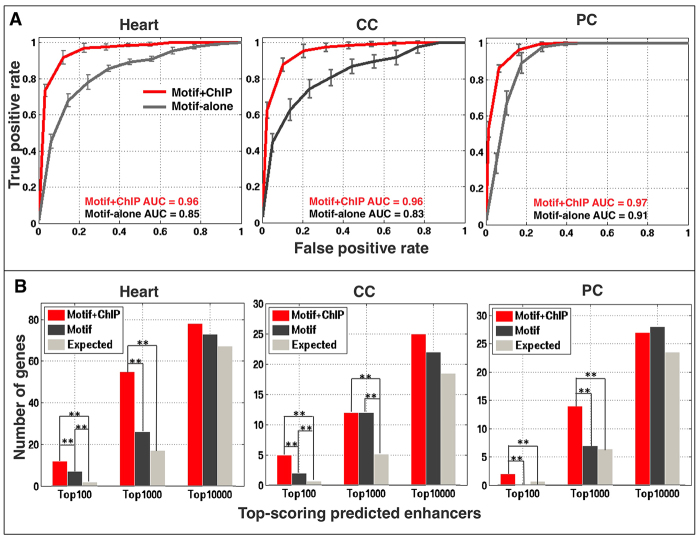
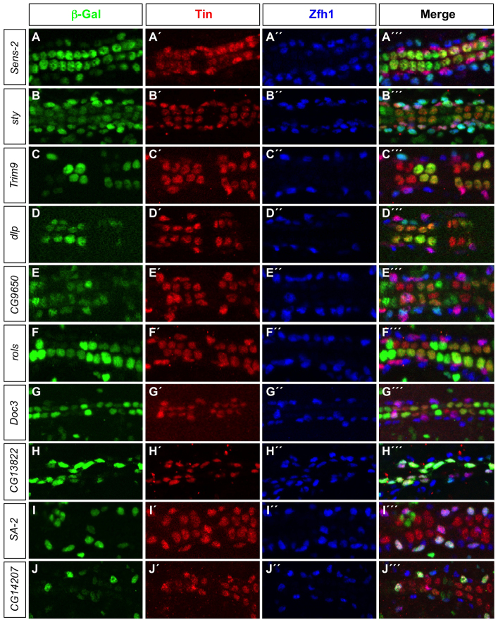
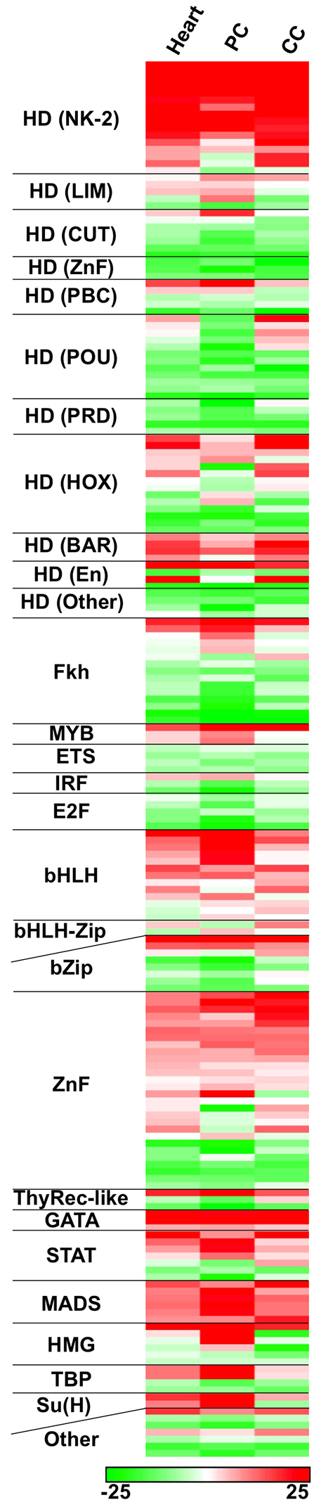
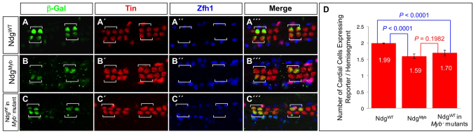
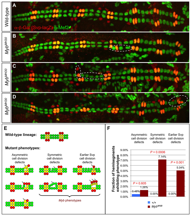
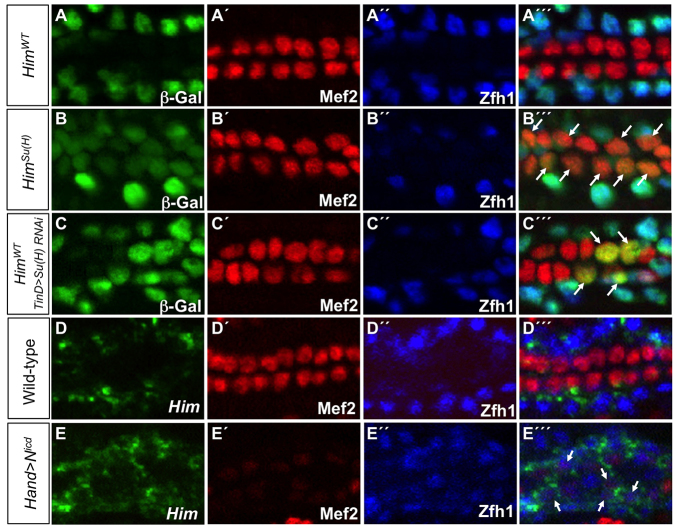
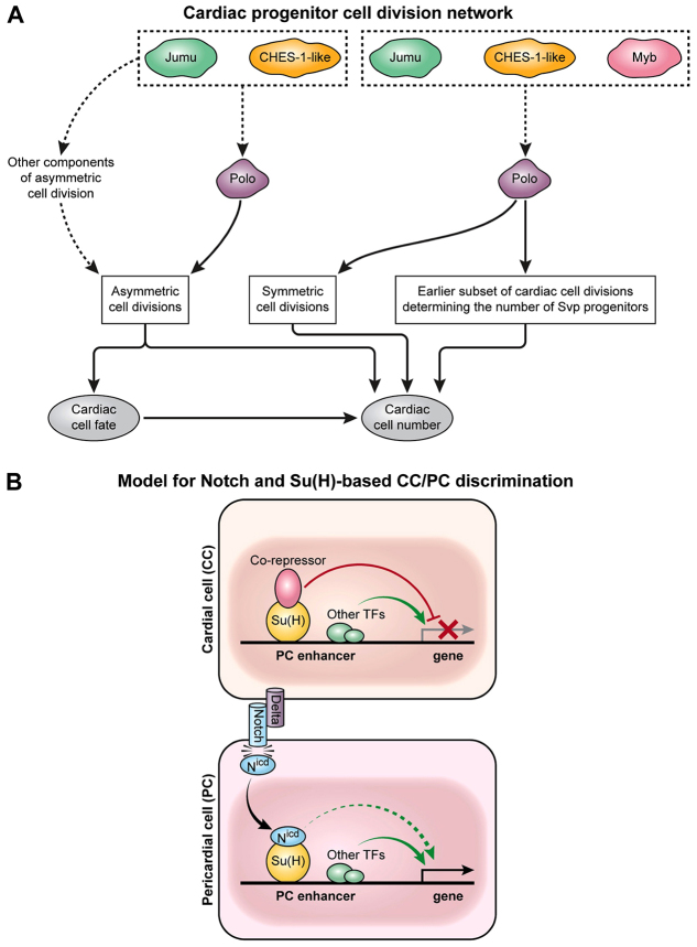
The Drosophila heart is composed of two distinct cell types, the contractile cardial cells (CCs) and the surrounding non-muscle pericardial cells (PCs), development of which is regulated by a network of conserved signaling molecules and transcription factors (TFs). Here, we used machine learning with array-based chromatin immunoprecipitation (ChIP) data and TF sequence motifs to computationally classify cell type-specific cardiac enhancers. Extensive testing of predicted enhancers at single-cell resolution revealed the added value of ChIP data for modeling cell type-specific activities. Furthermore, clustering the top-scoring classifier sequence features identified novel cardiac and cell type-specific regulatory motifs. For example, we found that the Myb motif learned by the classifier is crucial for CC activity, and the Myb TF acts in concert with two forkhead domain TFs and Polo kinase to regulate cardiac progenitor cell divisions. In addition, differential motif enrichment and cis-trans genetic studies revealed that the Notch signaling pathway TF Suppressor of Hairless [Su(H)] discriminates PC from CC enhancer activities. Collectively, these studies elucidate molecular pathways used in the regulatory decisions for proliferation and differentiation of cardiac progenitor cells, implicate Su(H) in regulating cell fate decisions of these progenitors, and document the utility of enhancer modeling in uncovering developmental regulatory subnetworks.

Keywords: Cell division; Drosophila; Gene regulation; Machine learning; Organogenesis; Progenitor specification; Transcription factors.


Figures

Fig. 1.
Fig. 1.
Schematic overview of the computational and experimental strategy utilized in this study. Training sets of enhancers expressed in different cardiac cell types (increased in size through phylogenetic profiling) and control sequences were compiled and subsequently scanned to map sequence features corresponding to known binding site motifs collected from public databases and to in vivo TF binding signals obtained from published ChIP data profiles. Classifiers were built to create enhancer models that discriminate cell type-specific enhancers from respective controls. For each cell type, two classifiers were independently constructed: one based solely on motif features (‘motif-alone’) and the other on motif features and ChIP signals (‘motif+ChIP′). The enhancer models were used to scan the Drosophila genome to identify novel cell-specific enhancers similar to the training sets, and the reliability and efficacy of the motif-alone and motif+ChIP classifiers for different cardiac cell types were examined and compared. Sequence features positively associated with computational classification were examined further using cis and trans in vivo experimental assays to identify and determine the functional roles of both the binding motifs and their associated regulatory TFs.
Fig. 2.
Fig. 2.
Cell-specific cardiac enhancer classifiers perform with high specificity and sensitivity. (A) Average receiver operating characteristic curves and standard deviations for tenfold cross-validation performed for both motif-alone and motif+ChIP classifiers in 20 independent runs. (B) Enrichment in validated heart, CC and PC genes in the neighborhood of putative cardiac enhancers at different ranks for the motif-alone and motif+ChIP classifiers. Each of these genes is generally associated with only one prediction. Double asterisks indicate significant differences (P<0.005).
Fig. 3.
Fig. 3.
Candidate enhancers predicted by the cell-specific cardiac classifiers are active in the appropriate cardiac cell types. (A-J′′) lacZ reporter gene activity (β-galactosidase, green) driven by classifier-predicted enhancers. All PCs are marked by Zfh1 expression (blue) whereas the posterior-most four CCs in each hemisegment, the Tin-PCs and the Eve-PCs are marked by Tin expression (red).
Fig. 4.
Fig. 4.
Machine learning modeling of cardiac enhancers reveals sequence motif features that are relevant to their cell type-specific functional classification. A linear SVM was trained for each of three cardiac training sets (heart, PC and CC) using motif features and relevant ChIP data. TF binding motifs are ranked according to their linear SVM weights, with positive weights reflecting enrichment and negative weights reflecting depletion from the training set compared with background. TF binding motifs were grouped according to DNA-binding domain class of their respective TFs.
Fig. 5.
Fig. 5.
Myb is an activator of the Ndg enhancer in the heart. (A-C′′) The posterior-most four CCs are marked by Tin expression (red), and the PCs are marked by Zfh1 expression (blue). (A-A′′) A β-galactosidase reporter (green) driven by the wild-type Ndg enhancer is expressed in only two Tin-expressing CCs per hemisegment (square brackets). (B-B′′) Mutations in the Myb binding sites result in partial but significant inactivation of reporter expression in these two CCs. (C-C′′) Reporter expression from the wild-type Ndg enhancer is also partially but significantly inactivated in these two CCs in embryos hemizygous for the MybMH30 null mutation. (D) Histogram showing the mean number of CCs with 95% confidence intervals expressing the reporter and the significance of partial inactivation as a result of either the Myb binding site mutations in the Ndg enhancer or the MybMH30 null mutation.
Fig. 6.
Fig. 6.
Cardiac progenitor cell division defects associated with Myb loss of function. (A) A heart from a wild-type embryo bearing the svp-lacZ enhancer trap showing hemisegments consisting of four Tin-CCs (green), two Svp-CCs (yellow) and two Svp-PCs (red). (B-D) Hearts from embryos that are hemizygous for the MybMH30 null mutation demonstrating mutant hemisegments with either excess or too few CCs (dashed ovals) and illustrating the two distinct types of progenitor cell division defects that underlie these cardiac phenotypes. (E) Schematic showing cell lineage relationships in a wild-type heart, and the three previously characterized cardiac progenitor cell divisions defects known to be responsible for localized changes in heart cell number (Ahmad et al., 2012). Note that only two types of developmental errors, those involved in symmetric cell divisions and those involved in an earlier step to determine the number of Svp progenitors, are primarily responsible for the Myb cardiac phenotypes. (F) Fraction of hemisegments exhibiting each type of cardiac progenitor cell division defect in embryos that are wild type or hemizygous for the MybMH30 mutation. The significance of each type of cell division defect in the Myb mutants compared with wild-type embryos is shown.
Fig. 7.
Fig. 7.
Su(H) discriminates between PC and CC enhancer activities. (A-C′′) lacZ reporter gene activity (β-galactosidase, green) driven by relevant Him enhancers in indicated genotypes. All CCs express Mef2 (red) whereas PCs are marked by Zfh1 (blue). (A-A′′) The wild-type Him enhancer (HimWT) is active only in the Zfh1-expressing PCs. (B-B′′) When the Su(H) binding site is mutated in the Him enhancer [HimSu(H)], the reporter is still active in Zfh1-expressing PCs but is de-repressed in Mef2-positive CCs (arrows). (C-C′′) Knockdown of Su(H) with dorsal mesoderm-targeted RNAi driven by the TinD-GAL4 driver induces ectopic HimWT enhancer-driven β-galactosidase reporter activity in CCs (arrows). (D-E′′) Fluorescent in situ hybridization analysis of stage 16 embryos for Him mRNA (D,E) and antibody analysis for Mef2 (D′,E′) and Zfh1 (D′,E′) of the indicated genotypes. (D-D′′) Him mRNA is restricted to the Zfh1-expressing PCs of the wild-type heart. (E-E′′) Overexpression of Nicd in all cells of the heart driven by the Hand-GAL4 driver leads to ectopic Him mRNA being expressed in the weakly Mef2-expressing CCs (arrows).
Fig. 8.
Fig. 8.
Distinct developmental subnetworks regulate cardiac progenitor cell division and specification. (A) Schematic of Jumu, CHES-1-like and Myb regulation of cardiac progenitor cell divisions by Polo kinase. The forkhead TFs Jumu and CHES-1-like regulate Polo kinase activity to mediate three distinct classes of cardiac progenitor cell divisions: asymmetric cell divisions, symmetric cell divisions, and an earlier round of cell division that determines the number of Svp progenitors (Ahmad et al., 2012). Mutations in Myb result in defects in only the latter two categories of cell divisions, which also exhibit synergistic genetic interactions among Myb, jumu, CHES-1-like and polo. As Myb transcriptionally regulates polo (Wen et al., 2008), Myb and the forkhead TFs act in concert to control Polo activity and thus govern both the symmetric and earlier class of cell divisions. (B) Schematic of the involvement of the Notch signaling pathway in the lineage decision between PCs and CCs for PC enhancers like that of Him. Modes of regulation activating and repressing target genes are shown as green and red arrows, respectively. In CCs, the enhancers of PC genes are repressed by the Su(H)-co-repressor complex. The Delta ligand expressed by CCs activates Notch receptor in neighboring PCs, with the resulting cleaved Nicd fragment associating with Su(H) and displacing the co-repressor. The consequent elimination of repressor complex binding in PCs is sufficient to initiate transcription due to the presence of other local TF activators, and is enhanced further by the Nicd-Su(H) complex.

Similar articles

Cited by

References

    1. Ahmad S. M., Tansey T. R., Busser B. W., Nolte M. T., Jeffries N., Gisselbrecht S. S., Rusan N. M., Michelson A. M. (2012). Two forkhead transcription factors regulate the division of cardiac progenitor cells by a Polo-dependent pathway. Dev. Cell 23, 97–111 - PMC - PubMed
    1. Bailey T. L., Gribskov M. (1998). Combining evidence using p-values: application to sequence homology searches. Bioinformatics 14, 48–54 - PubMed
    1. Barolo S., Posakony J. W. (2002). Three habits of highly effective signaling pathways: principles of transcriptional control by developmental cell signaling. Genes Dev. 16, 1167–1181 - PubMed
    1. Berger M. F., Philippakis A. A., Qureshi A. M., He F. S., Estep P. W., III, Bulyk M. L. (2006). Compact, universal DNA microarrays to comprehensively determine transcription-factor binding site specificities. Nat. Biotechnol. 24, 1429–1435 - PMC - PubMed
    1. Bernard F., Krejci A., Housden B., Adryan B., Bray S. J. (2010). Specificity of Notch pathway activation: twist controls the transcriptional output in adult muscle progenitors. Development 137, 2633–2642 - PMC - PubMed

Publication types

MeSH terms

Substances

LinkOut - more resources