. 2014 Feb;141(4):889-98.
doi: 10.1242/dev.102152.

Macroglobulin complement-related encodes a protein required for septate junction organization and paracellular barrier function in Drosophila

Affiliations

Macroglobulin complement-related encodes a protein required for septate junction organization and paracellular barrier function in Drosophila

Sonia Hall et al. Development. 2014 Feb.

Abstract

Polarized epithelia play crucial roles as barriers to the outside environment and enable the formation of specialized compartments for organs to carry out essential functions. Barrier functions are mediated by cellular junctions that line the lateral plasma membrane between cells, principally tight junctions in vertebrates and septate junctions (SJs) in invertebrates. Over the last two decades, more than 20 genes have been identified that function in SJ biogenesis in Drosophila, including those that encode core structural components of the junction such as Neurexin IV, Coracle and several claudins, as well as proteins that facilitate the trafficking of SJ proteins during their assembly. Here we demonstrate that Macroglobulin complement-related (Mcr), a gene previously implicated in innate immunity, plays an essential role during embryonic development in SJ organization and function. We show that Mcr colocalizes with other SJ proteins in mature ectodermally derived epithelial cells, that it shows interdependence with other SJ proteins for SJ localization, and that Mcr mutant epithelia fail to form an effective paracellular barrier. Tissue-specific RNA interference further demonstrates that Mcr is required cell-autonomously for SJ organization. Finally, we show a unique interdependence between Mcr and Nrg for SJ localization that provides new insights into the organization of the SJ. Together, these studies demonstrate that Mcr is a core component of epithelial SJs and also highlight an interesting relationship between innate immunity and epithelial barrier functions.

Keywords: Epithelia; Innate immunity; Septate junction.


Figures

Fig. 1.
Fig. 1.
Mutations in Mcr are embryonic lethal with phenotypes associated with SJ defects. (A) Schematic of the Mcr gene and Mcr protein. The insertion site of McrEY07421 and the amino acid substitution in Mcr1 are shown. The region of Mcr used for antibody generation is indicated by a bracket. ss, signal sequence; MG1, alpha-2-macroglobulin MG1 domain; A2M_N, MG2 domain; A2M_N_2, alpha-2-macroglobulin family N-terminal region; LDLa, low-density lipoprotein receptor A domain; A2M, alpha-2-macroglobulin family; A2M_comp, alpha-2-macroglobulin complement component; A2M_rec, alpha-2-macroglobulin receptor; TM, predicted transmembrane domain. (B-E) Cuticle preparations of a w1118 (wild-type) late embryo (B) and McrEY07421 (C), Mcr1 (D) and da-GAL4>UAS-Mcr-RNAi (E) mutant embryos. Anterior is left and dorsal is up or facing. Note the dorsal hole in the Mcr1 embryo (asterisk in D) and the ectopic salivary gland deposition in mutant animals (arrows in C-E). (F,G) Confocal optical sections of stage 17 w1118 (F) and Mcr1 mutant (G) embryos stained with Rhodamine-labeled wheat germ agglutinin (WGA), showing a highly convoluted trachea (arrows) in the Mcr1 animal. Scale bars: 100 μm.
Fig. 2.
Fig. 2.
Mcr is required for SJ structure and paracellular barrier function. (A-O) Confocal optical sections of salivary glands (A,D,G,J,M) stained with antibodies against Crb (red, and in A′,D′,G′,J′,M′) and Cor (green, and in A′,D′,G′,J′,M′), and hindguts (B,E,H,K,N) and trachea (C,F,I,L,O) from stage 16 w1118 (A-C), Df(2L)Exel7034 (D-F), Mcr1 (G-I), McrEY07421 (J-L) and McrPex3 (M-O) embryos. The salivary gland in G is from the same animal as the hindgut in H, as are the salivary gland and hindgut shown in J and K. Apical surfaces face the lumen of the gland. The wild-type salivary gland epithelium is outlined by a dashed line in A. Note that Cor is mislocalized along the lateral membrane in Mcr mutant salivary glands (arrows), whereas Crb is unaffected. (P-Q′) Differential interference contrast (P,Q) and fluorescence (P′,Q′) photomicrographs of the tracheal dorsal trunk in stage 17 w1118 (P) and Mcr1 (Q) embryos that had been injected with a 10 kDa Rhodamine-labeled dextran. The labeled dextran does not cross the tracheal epithelium and enter the lumen in the wild-type embryo, but does in the Mcr1 mutant embryo (arrows). Scale bars: 20 μm.
Fig. 3.
Fig. 3.
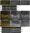
Mcr transcript expression and Mcr protein expression and localization during development. (A) Northern blot analysis of total RNA isolated from staged w1118 embryos probed for Mcr expression. Numbers refer to hours after egg laying. Hybridization to rp49 (RpL32) was used as a control for loading and transfer. (B-J) Confocal optical sections of w1118 embryos (B-E), w1118 third instar larval wing imaginal disc (F-H), He-GAL4, UAS GFP third instar larval hemocyte (I) and w1118 ovariole (J) stained with antibodies against Mcr alone (B-D), or co-stained with antibodies against Mcr (red, and in E′-J′) and Cor (green, and in E′-H′), GFP (green in I) or Fas3 (green in J, and in J′). (B-E) Mcr is associated with the membrane in stage 11 embryos (B) and by stage 13 is obviously expressed in ectodermally derived epithelia including the epidermis (ep), foregut (fg), hindgut (hg), salivary gland (sg) and trachea (tr) (C). In stage 14 embryos (D), Mcr is enriched at the apical lateral region of the membrane, but is also expressed more basolaterally (arrow). By stage 16 (E; in the hindgut), Mcr colocalizes with Cor in the region of the SJ. In third instar wing imaginal discs (F-H), Mcr colocalizes with Cor in the apical region of the lateral membrane of the disc proper cells, which can be seen in deeper sections (G) where lateral membranes lie adjacent to the folds in the epithelium (arrow), and by rendering an xz transverse section from a confocal z-series (H). A higher magnification view (outset in H) shows that Cor localization extends more basally than Mcr (arrows), and that Mcr is also expressed on the apical surface in a domain independent of Cor (arrowhead; note that staining in the peripodial epithelium can be seen above this line). (I) Confocal optical section of a He-GAL4, UAS-GFP hemocyte stained with antibodies against Mcr (red) and GFP (green) and with DAPI (blue). Mcr is expressed in larval hemocytes, but is largely found inside the cell. (J) In the ovary, Mcr is most strongly expressed in stage 1 of the germarium (arrow) and in polar follicle cells (arrowhead), where it colocalizes with Fas3. Mcr is also expressed at lower levels in the follicle cells and at the membrane in the germ cells. Scale bars: 20 μm.
Fig. 4.
Fig. 4.
Mcr is mislocalized in the hindguts of SJ mutant embryos. (A-G) Confocal optical sections of hindguts (A-E) and salivary glands (F,G) from stage 16 w1118 (A), Nrx-IV4304 (B), nrvZCL164 (C), crokKG6053 (D,G), Tsf2KG01571 (E) and sinunwu7 (F) embryos stained with antibodies against Mcr (red, and in A′-G′) and Cor (green, and in A′-G′). Mcr is mislocalized along the basolateral domain in the hindgut epithelia coincident with Cor in all of these embryos (arrows), and also shows strong mislocalization on the apical plasma membrane of Tsf2KG01571 embryos (E) that is independent of Cor (asterisks). Scale bars: 20 μm. (H) Plot of average fluorescence recovery after photobleaching (FRAP) of Nrx-IV-GFP in the epidermis of stage 15 wild-type (gray line) and McrEY07421 mutant (black squares) animals. Error bars indicate s.e.m.
Fig. 5.
Fig. 5.
Mcr is required cell-autonomously for SJ organization in embryonic epithelia and larval wing imaginal discs. (A,B) Confocal optical sections (A) and xz rendering from a confocal z-series (B) of the epidermis of a stage 16 UAS-Dcr-2; en-GAL4/UAS-Mcr-RNAi embryo stained with antibodies against Mcr (red, and in A′,B′) and Cor (green, and in A′,B′). The xz rendering is along the white line in A. Mcr expression is strongly reduced in the posterior cells, although the majority of the remaining protein appears to be membrane associated (arrow in A′, compared with Cor localization in A′). Note that Cor is not as enriched in the apical regions of Mcr-RNAi cells, indicating a disorganization of the SJ junction (B). (C-E) Confocal optical section and xz rendering from a confocal z-series from a late third instar UAS-Dcr-2; ap-GAL4/UAS-Mcr-RNAi wing imaginal disc stained with antibodies against Mcr (red, and in C′-E′) and Cor (green, and in C′-E′). Dorsal is to the top in C and D and to the left in E, and the dorsal-ventral boundary is indicated by arrows in C. D is a higher magnification of the boxed region in C. Mcr is strongly reduced in the dorsal compartment of Mcr-RNAi wing discs, but the residual Mcr appears to align with Cor at the boundary between adjacent cells (arrows in D) and is apically enriched in the lateral membrane (arrows in E). Wild-type cells at the dorsal-ventral boundary have reduced Mcr expression along the membrane in contact with Mcr-RNAi cells (red arrowheads in D′). Scale bars: 20 μm.
Fig. 6.
Fig. 6.
The SJ localization of Mcr and of Nrg depend upon each other. (A-C) Confocal optical section and xz rendering from a confocal z-series of a third instar UAS-Dcr-2; ap-GAL4/UAS-Nrg-RNAi wing imaginal disc stained with antibodies against Mcr (red, and in A′-C′) and Cor (green, and in A′-C′). Dorsal is to the top in A and B and to the left in C, and the dorsal-ventral boundary is indicated by arrows in A. (B) Higher magnification of the boxed region in A. Mcr is strongly reduced in Nrg-RNAi cells, but appears to colocalize with Cor along the boundary between adjacent cells (arrows in B), and is apically enriched in the lateral membrane (arrow in C′). Note that the apical plasma membrane expression of Mcr is also eliminated in the Nrg-RNAi cells (red arrowheads). (D) Confocal optical section of the hindgut from a stage 16 Nrg17 embryo stained with antibodies against Mcr (red, and in D′) and Cor (green, and in D′). Whereas Cor is mislocalized along the lateral membrane in the hindgut of the Nrg17 embryo, Mcr is nearly absent from the lateral membrane. (E-F′) Confocal optical sections of a stage 11 (E) and stage 15 (F) Nrg17 embryo stained with antibodies against Mcr. A higher magnification view of the salivary gland in F is shown in F′. Note that Mcr is expressed and shows some membrane localization in the stage 11 Nrg embryo (arrows in E), but is largely lost from the lateral membrane at the expense of the apical plasma membrane by stage 15 (arrows). (G,H) Confocal optical sections of salivary glands from a stage 16 McrEY07421 heterozygous (G) and homozygous (H) animal stained with antibodies against Mcr (red) and Nrg (green, and in H′). Mcr and Nrg colocalize at the SJ in the heterozygous animal (arrow in G), whereas Nrg predominantly localizes to the apical surface in the Mcr mutant animal (arrow in H′). Scale bars: 20 μm.

Similar articles

Cited by

References

    1. Armstrong P. B., Quigley J. P. (1999). Alpha2-macroglobulin: an evolutionarily conserved arm of the innate immune system. Dev. Comp. Immunol. 23, 375–390 - PubMed
    1. Baumgartner S., Littleton J. T., Broadie K., Bhat M. A., Harbecke R., Lengyel J. A., Chiquet-Ehrismann R., Prokop A., Bellen H. J. (1996). A Drosophila neurexin is required for septate junction and blood-nerve barrier formation and function. Cell 87, 1059–1068 - PubMed
    1. Behr M., Riedel D., Schuh R. (2003). The claudin-like megatrachea is essential in septate junctions for the epithelial barrier function in Drosophila. Dev. Cell 5, 611–620 - PubMed
    1. Bieber A. J., Snow P. M., Hortsch M., Patel N. H., Jacobs J. R., Traquina Z. R., Schilling J., Goodman C. S. (1989). Drosophila neuroglian: a member of the immunoglobulin superfamily with extensive homology to the vertebrate neural adhesion molecule L1. Cell 59, 447–460 - PubMed
    1. Bou Aoun R., Hetru C., Troxler L., Doucet D., Ferrandon D., Matt N. (2011). Analysis of thioester-containing proteins during the innate immune response of Drosophila melanogaster. J. Innate Immun. 3, 52–64 - PMC - PubMed

Publication types

MeSH terms

LinkOut - more resources