. 2014 Feb;141(4):962-73.
doi: 10.1242/dev.098327.

Proteomic profiling of cardiac tissue by isolation of nuclei tagged in specific cell types (INTACT)

Affiliations

Proteomic profiling of cardiac tissue by isolation of nuclei tagged in specific cell types (INTACT)

Nirav M Amin et al. Development. 2014 Feb.

Abstract

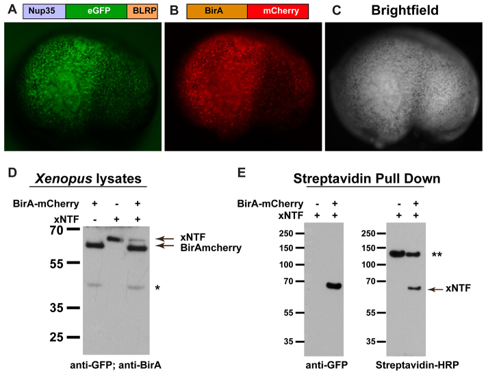
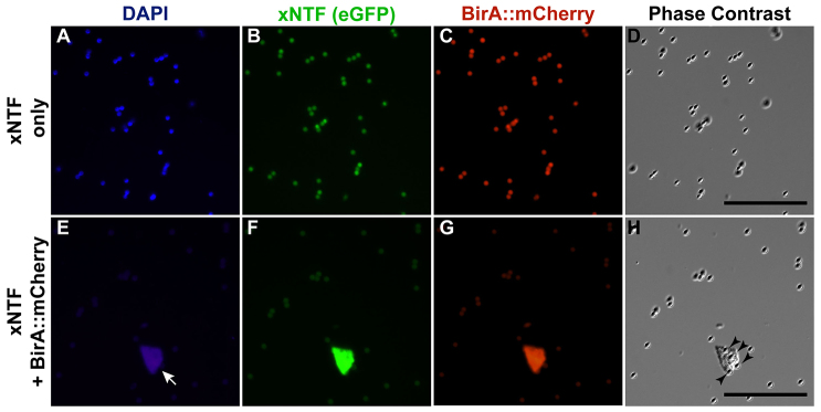
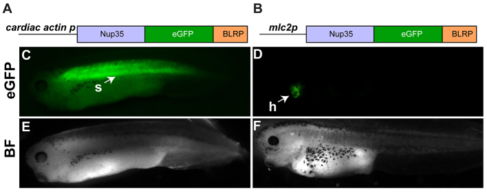
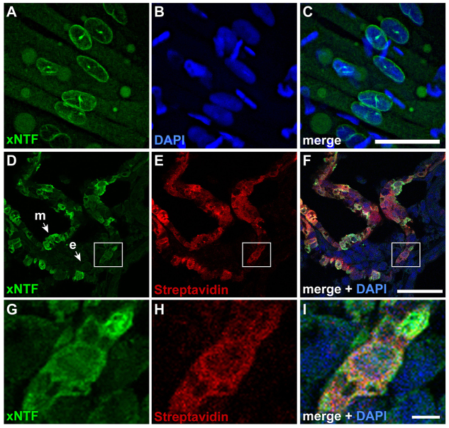
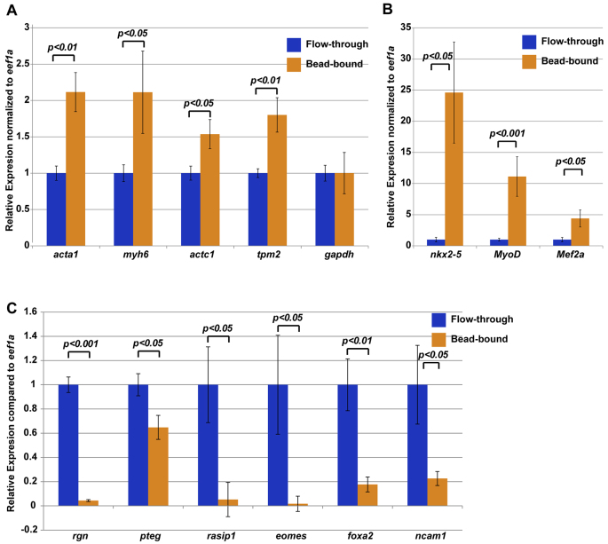
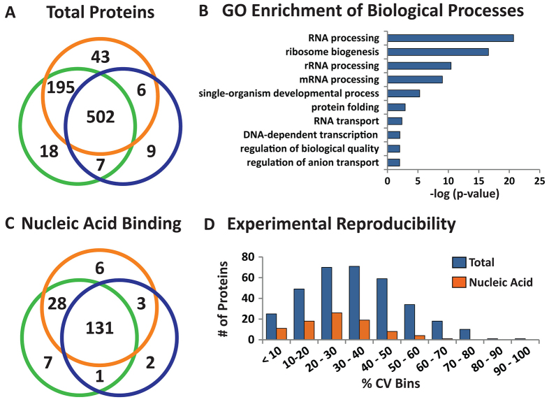
The proper dissection of the molecular mechanisms governing the specification and differentiation of specific cell types requires isolation of pure cell populations from heterogeneous tissues and whole organisms. Here, we describe a method for purification of nuclei from defined cell or tissue types in vertebrate embryos using INTACT (isolation of nuclei tagged in specific cell types). This method, previously developed in plants, flies and worms, utilizes in vivo tagging of the nuclear envelope with biotin and the subsequent affinity purification of the labeled nuclei. In this study we successfully purified nuclei of cardiac and skeletal muscle from Xenopus using this strategy. We went on to demonstrate the utility of this approach by coupling the INTACT approach with liquid chromatography-tandem mass spectrometry (LC-MS/MS) proteomic methodologies to profile proteins expressed in the nuclei of developing hearts. From these studies we have identified the Xenopus orthologs of 12 human proteins encoded by genes, which when mutated in human lead to congenital heart disease. Thus, by combining these technologies we are able to identify tissue-specific proteins that are expressed and required for normal vertebrate organ development.

Keywords: Cardiac muscle; INTACT; Proteomics; Skeletal muscle; Xenopus.


Figures

Fig. 1.
Fig. 1.
Affinity isolation of biotin-tagged proteins from Xenopus embryos. (A-C) Schematic of Nup35::eGFP::BLRP(xNTF) and BirA::mCherry fusion proteins, and corresponding images of a Xenopus embryo co-injected at the one-cell stage with capped mRNA coding for each fusion. Lateral view of a stage 23 embryo with anterior to the left and dorsal up. (D) Western blot analysis of embryos injected individually and together with xNTF and BirA::mCherry shows that the proteins are of the correct size. xNTF was detected with anti-GFP antibodies, and BirA was detected by anti-BirA antibodies. (E) Immunoprecipitation of xNTF with streptavidin-conjugated beads, followed by western blot analysis shows that xNTF is efficiently isolated from embryos by probing with anti-GFP or streptavidin-HRP. *A potential translation product of BirA::mCherry corresponding to the size of BirA protein alone. **Endogenously biotinylated proteins isolated by streptavidin.
Fig. 2.
Fig. 2.
Affinity isolation of xNTF-tagged nuclei. Streptavidin-coated magnetic beads incubated with nuclei from embryos injected with mRNA encoding xNTF with or without BirA::mCherry. (A-D) Fluorescent DAPI (A), xNTF (eGFP; B), BirA::mCherry (Texas Red; C) and phase-contrast (D) images of magnetic beads after nuclear purification of xNTF in the absence of BirA. Autofluorescing beads are detectable throughout the field, but no nuclei. (E-H) Fluorescent DAPI (E), eGFP (F), Texas Red (G) and phase-contrast (H) images of magnetic beads after nuclear purification of xNTF. Note the presence of eGFP and BirA::mCherry-positive nucleus (arrow) and presence of magnetic beads coating nucleus (arrowheads). Scale bars: 50 μm.
Fig. 3.
Fig. 3.
Generation of transgenic embryos expressing xNTF. (A,B) Schematics of constructs used to generate transgenic embryos expressing xNTF in the striated muscle and myocardium (A) and the myocardium only (B). (C-F) Representative images of stage 38 (C,E) and stage 40 (D,F) embryos harboring the respective transgenes. Embryos express xNTF (eGFP) in the expected pattern (C,D), and appear morphologically normal (E,F). eGFP in myocardium is fainter than staining in somites and masked by autofluorescence of the gut in C. Images shown are lateral views with anterior to the left and dorsal up. BF, bright field; h, heart; s, somites.
Fig. 4.
Fig. 4.
Demonstration of xNTF localization and biotin tagging in transgenic animals. (A-C) xNTF localization in the somites as detected by eGFP (A); nuclei were marked with DAPI (B) and the merged image is shown in C. Images are from whole embryos with anterior to the left and dorsal up. (D-F) Transverse sections through the heart (dorsal is up) show expression of xNTF (D) in the myocardium (m) but not the endocardium (e), and corresponding localization of biotinylated xNTF as detected by streptavidin (E). Images were overlaid with DAPI to show the nuclei in F. (G-I) Magnified image of the boxed region in D-F. Scale bars: 200 μm (C); 50 μm (F); 5 μm (I).
Fig. 5.
Fig. 5.
INTACT purified nuclei are enriched for cardiac and/or skeletal muscle markers. (A,B) qRT-PCR results show that skeletal muscle structural genes (A) and cardiac or skeletal transcription factors (B) are enriched in bead-bound nuclei compared with flow-through of total nuclei. All expression levels are relative to eef1a and flow-through was normalized to one. Expression of gapdh, another housekeeping gene, was unchanged in bead-bound nuclei compared with flow-through nuclei. (C) qRT-PCR demonstrates that INTACT nuclei show reduced levels of gene expression of non-muscle genes. Error bars represent s.e.m. from a single experiment. Three biological replicates of nuclear purifications were performed. P-values from Student’s t-tests are indicated.
Fig. 6.
Fig. 6.
Proteomic profiling of INTACT-enriched Xenopus cardiomyocyte nuclei. (A) Venn diagram comparison of the total proteins identified in three biological replicates (n=781). (B) g:profiler biological process gene ontology enrichment analysis. Only terms that were significant versus the annotated Xenopus tropicalis genome are shown (-log(P-value) >2). (C) Venn diagram comparison of proteins identified with ‘nucleic acid binding’ molecular function gene ontology (n=178). (D) Distribution of weighted spectral count percentage coefficient of variation (% CV, n=3). % CV was calculated for shared proteins identified in A and C, corresponding to total proteins (blue bars) and nucleic acid-binding proteins (orange bars) with at least three weighted spectral counts in all biological replicates.

Similar articles

Cited by

References

    1. Afouda B. A., Hoppler S. (2009). Xenopus explants as an experimental model system for studying heart development. Trends Cardiovasc. Med. 19, 220–226 - PubMed
    1. Agostini M., Tucci P., Killick R., Candi E., Sayan B. S., Rivetti di Val Cervo P., Nicotera P., McKeon F., Knight R. A., Mak T. W., et al. (2011). Neuronal differentiation by TAp73 is mediated by microRNA-34a regulation of synaptic protein targets. Proc. Natl. Acad. Sci. USA 108, 21093–21098 - PMC - PubMed
    1. Ahmed R. P., Haider H. K., Buccini S., Li L., Jiang S., Ashraf M. (2011). Reprogramming of skeletal myoblasts for induction of pluripotency for tumor-free cardiomyogenesis in the infarcted heart. Circ. Res. 109, 60–70 - PMC - PubMed
    1. Al-Saaidi R., Rasmussen T. B., Palmfeldt J., Nissen P. H., Beqqali A., Hansen J., Pinto Y. M., Boesen T., Mogensen J., Bross P. (2013). The LMNA mutation p.Arg321Ter associated with dilated cardiomyopathy leads to reduced expression and a skewed ratio of lamin A and lamin C proteins. Exp. Cell Res. 319, 3010–3019 - PubMed
    1. Amaya E., Kroll K. L. (1999). A method for generating transgenic frog embryos. Methods Mol. Biol. 97, 393–414 - PubMed

Publication types