. 2014 Feb 18;111(7):2698-703.
doi: 10.1073/pnas.1320243111. Epub 2014 Feb 4.

Transcriptional activation of hypoxia-inducible factor-1 (HIF-1) in myeloid cells promotes angiogenesis through VEGF and S100A8

Affiliations

Transcriptional activation of hypoxia-inducible factor-1 (HIF-1) in myeloid cells promotes angiogenesis through VEGF and S100A8

G-One Ahn et al. Proc Natl Acad Sci U S A. .

Abstract

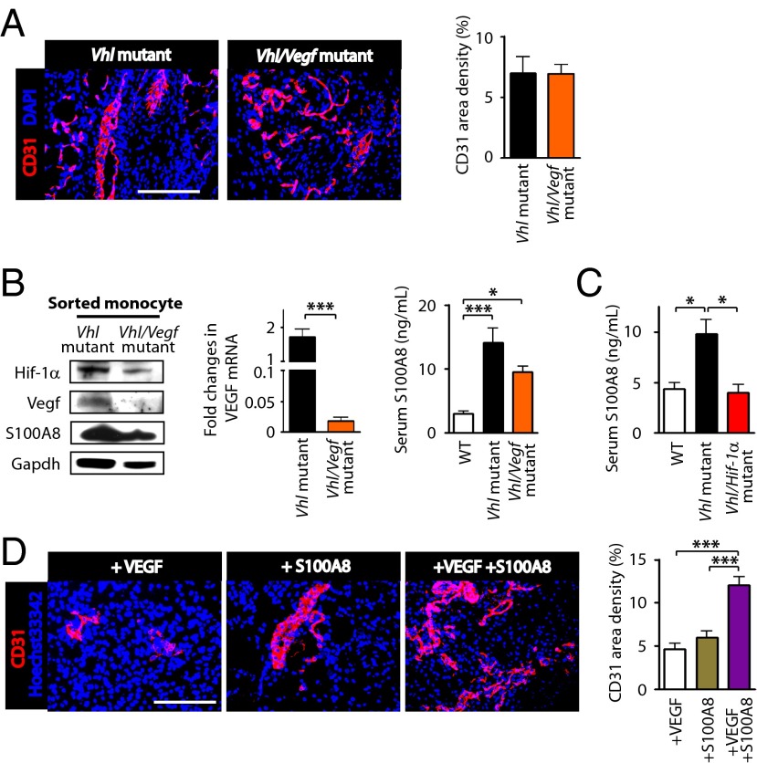
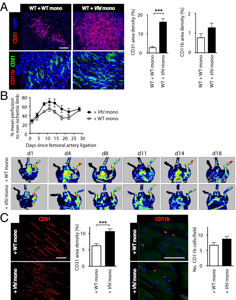
Emerging evidence indicates that myeloid cells are essential for promoting new blood vessel formation by secreting various angiogenic factors. Given that hypoxia-inducible factor (HIF) is a critical regulator for angiogenesis, we questioned whether HIF in myeloid cells also plays a role in promoting angiogenesis. To address this question, we generated a unique strain of myeloid-specific knockout mice targeting HIF pathways using human S100A8 as a myeloid-specific promoter. We observed that mutant mice where HIF-1 is transcriptionally activated in myeloid cells (by deletion of the von Hippel-Lindau gene) resulted in erythema, enhanced neovascularization in matrigel plugs, and increased production of vascular endothelial growth factor (VEGF) in the bone marrow, all of which were completely abrogated by either genetic or pharmacological inactivation of HIF-1. We further found that monocytes were the major effector producing VEGF and S100A8 proteins driving neovascularization in matrigel. Moreover, by using a mouse model of hindlimb ischemia we observed significantly improved blood flow in mice intramuscularly injected with HIF-1-activated monocytes. This study therefore demonstrates that HIF-1 activation in myeloid cells promotes angiogenesis through VEGF and S100A8 and that this may become an attractive therapeutic strategy to treat diseases with vascular defects.


Conflict of interest statement

The authors declare no conflict of interest.

Figures

Fig. 1.
Fig. 1.
Enhanced angiogenic phenotypes in mice deficient for pVHL in hS100A8 myeloid cells. (A) Vhl mutant mice exhibited erythema (black arrowheads) compared with WT mice. (B) Red blood cell (RBC) or hemoglobin (HGB) levels in WT (n = 4) or Vhl mutant (n = 10) mice. Data are the mean ± SEM. (C) Antibody array analyses of the Vhl mutant or WT mice using bone marrow lysates. Note that Vegf levels (red arrowhead) were increased approximately twofold in Vhl mutant mice. Quantitative results are shown in Table S1. (D) Immunostaining of matrigel implanted in WT or Vhl mutant mice for CD31 endothelial cells (red). Hoechst 33342 (blue) was i.v. administered immediately before the matrigel harvest. (Scale bar: 100 µm.) Quantification of CD31 area density is shown in the bar graph. Data are the mean ± SEM (n = 7 for WT; n= 18 for Vhl mutant mice). *P < 0.05 determined by unpaired Student t test. (E) Western blot of matrigel lysates obtained from Vhl mutant or WT mice. (F) Immunostaining of matrigel in D for CD11b (red) or CD31 (green). (Scale bar and n numbers are as in D.)
Fig. 2.
Fig. 2.
Angiogenic phenotypes in mice deficient for pVHL in hS100A8 myeloid cells require HIF-1 activation. (A) Inactivation of Hif-1α in Vhl mutant mice (Vhl/Hif-1α mutant) suppressed erythema of Vhl single mutant mice (black arrowheads). (B) Antibody array analyses as in Fig. 1 for Vhl or the Vhl/Hif-1α mutant mice bone marrow lysates. Note that the increased Vegf in Vhl mutant mice is significantly reduced in Vhl/Hif-1α mutant mice (red arrowheads). (C) Serum VEGF measured by ELISA (n = 5 per group). (D) Immunostaining (Upper) and Western blots (Lower) of matrigel implanted in Vhl mutant mice, Vhl mutant mice treated with NSC 134754, a HIF-1 inhibitor, or in Vhl/Hif-1α mutant mice. (Scale bar: 100 µm.) Quantification of CD31 area density is shown in the bar graph (n = 10 for Vhl mutant; n = 7 for Vhl mutant + NSC; n = 6 for Vhl/Hif-1α mutant). (E) Immunostaining of matrigel implanted in WT, Hif-1α mutant, Hif-2α mutant, or Hif-1α /Hif-2α mutant mice. Quantification of CD31 area densities in matrigel is shown in the bar graph (n = 7 for Vhl littermate; n = 7 for Vhl mutant; n = 6 for Hif-1α littermate; n = 10 for Hif-1α mutant; n = 4 for Hif-2α littermate; n = 6 for Hif-2α mutant; n = 5 for Hif-1α/Hif-2α littermate; n = 6 for Hif-1α/Hif-2α mutant). In C, D, and E, data are the mean ± SEM and *P < 0.05, **P < 0.01, and ***P < 0.001 determined by one-way ANOVA.
Fig. 3.
Fig. 3.
Bone marrow-derived cells mediate neovascularization in matrigel. (A) A picture (Upper) and Western blots (Lower) of matrigel in WT mice reconstituted with WT bone marrow (WT + WT BMT, n = 8) or with Vhl mutant mice bone marrow (WT + Vhl BMT, n = 8). (B) Immunofluorescent staining of matrigels in WT + WT BMT, WT + Vhl BMT, or Vhl + WT BMT for CD31 or CD11b. Nuclei were counterstained by DAPI. (Scale bar: 100 µm.) Quantification of CD31 or CD11b area density is shown in the bar graphs. Data are the mean ± SEM with *P < 0.05 determined by one-way ANOVA (n = 10 for WT + WT BMT; n = 8 for WT + Vhl BMT; n = 5 for Vhl + WT BMT).
Fig. 4.
Fig. 4.
Monocytes are the major effector mediating angiogenic effects. (A) Immunophenotyping analyses of CMPs, GMPs, PreGs, granulocytes (Gr), and monocytes (Mono) of WT or Vhl mutant mice. (B) qRT-PCR analyses in 20,000 cells sorted from each myeloid subpopulation for Hif-1α, Vhl, and Vegf. Results are quantified as fold changes of mRNA in Vhl mutant over WT mice. *P < 0.05, **P < 0.01, and ***P < 0.001 determined by one-way ANOVA using triplicate determinations from pooled samples of two animals per group. Data are the mean ± SEM. (C) Western blots performed with FACS sorted monocytes obtained from WT, Vhl mutant, or Vhl/Hif-1α mutant mice.
Fig. 5.
Fig. 5.
VEGF and S100A8 act cooperatively to promote neovascularization in matrigel. (A) Matrigel immunostained for CD31 (red) implanted in Vhl mutant or Vhl/Vegf double mutant mice. Quantification of CD31 area densities are shown in the bar graph (n = 5 per group). (B) Western blot (Left) showing Hif-1α, Vegf, S100A8 protein levels in sorted monocytes obtained from Vhl or Vhl/Vegf mutant mice. qRT-PCR for Vegf (Center) was performed in these mice and quantified as fold changes compared with WT mice. ***P < 0.001 by unpaired Student t test. Serum S100A8 levels (Right) in WT, Vhl mutant, or Vhl/Vegf mutant mice. *P < 0.05 and ***P < 0.001, respectively, determined by one-way ANOVA (n = 6 per group). (C) Serum S100A8 measured in WT, Vhl mutant, or Vhl/Hif-1α mutant mice. *P < 0.05 (n = 5 per group). (D) Immunostaining of matrigel implanted in WT that had been admixed with VEGF alone, S100A8 alone, or VEGF in combination with S100A8. ***P < 0.001 determined by one-way ANOVA (n = 5 per group). (Scale bars: 100 µm in A and D.) Data in A–D are the mean ± SEM.
Fig. 6.
Fig. 6.
HIF-1–activated monocytes promote angiogenesis in matrigel and improve blood flow in a mouse model of hindlimb ischemia. (A) Immunostaining of matrigel implanted in WT mice that had been admixed with 100,000 monocytes isolated from WT (WT mono) or Vhl mutant (Vhl mono) mice for CD31 and CD11b. (Scale bar: 100 μm.) Area densities of CD31 and CD11b in matrigel are shown (Right). ***P < 0.001 determined by Student t test (n = 6 per group). (B) Laser Doppler flowmetry analysis of the blood perfusion in the femoral artery ligated WT animals injected intramuscularly with 50,000 monocytes isolated from WT mice (WT + WT mono) or Vhl mutant mice (WT + Vhl mono). The data are the mean ± SEM (n = 6 for WT + WT mono; n = 7 for WT + Vhl mono). Representative laser Doppler image from each group is shown (Lower). (C) Immunostaining of the quadrate muscle at day 14 for CD31 (Left) or CD11b (Right). ***P < 0.001 determined by unpaired Student t test. (Scale bars: 100 µm.) Data in AC are the mean ± SEM.

References

    1. Lamalice L, Le Boeuf F, Huot J. Endothelial cell migration during angiogenesis. Circ Res. 2007;100(6):782–794. - PubMed
    1. Granata F, et al. Production of vascular endothelial growth factors from human lung macrophages induced by group IIA and group X secreted phospholipases A2. J Immunol. 2010;184(9):5232–5241. - PMC - PubMed
    1. Medina RJ, et al. Myeloid angiogenic cells act as alternative M2 macrophages and modulate angiogenesis through interleukin-8. Mol Med. 2011;17(9-10):1045–1055. - PMC - PubMed
    1. Kujawski M, et al. Stat3 mediates myeloid cell-dependent tumor angiogenesis in mice. J Clin Invest. 2008;118(10):3367–3377. - PMC - PubMed
    1. Shojaei F, Zhong C, Wu X, Yu L, Ferrara N. Role of myeloid cells in tumor angiogenesis and growth. Trends Cell Biol. 2008;18(8):372–378. - PubMed

Publication types

MeSH terms