Review
. 2014 Feb;99(2):209-21.
doi: 10.3324/haematol.2013.087619.

Update on treatment of light chain amyloidosis

Affiliations
Review

Update on treatment of light chain amyloidosis

Shameem Mahmood et al. Haematologica. 2014 Feb.

Abstract

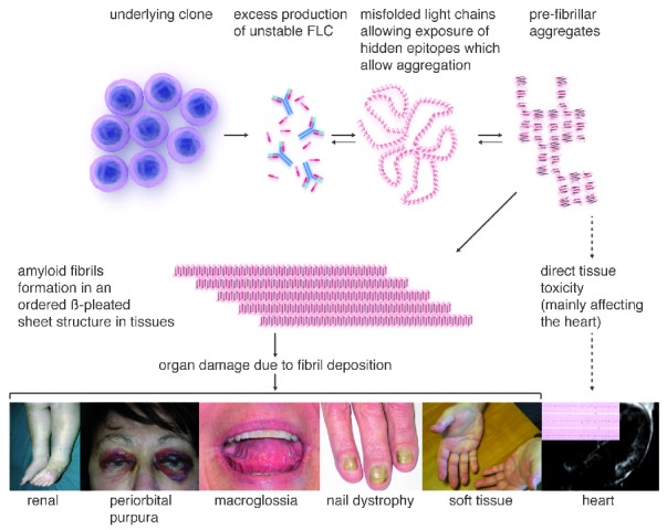
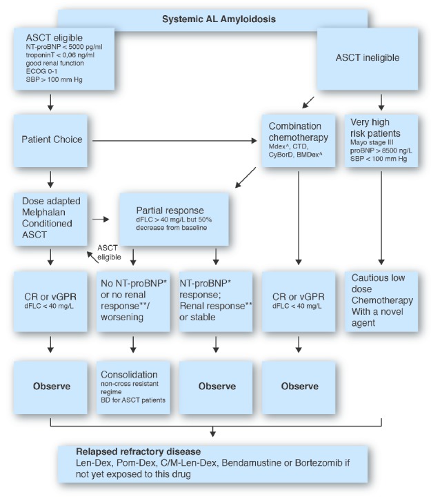
Light chain amyloidosis is the most common type of amyloidosis as a consequence of protein misfolding of aggregates composed of amyloid fibrils. The clinical features are dependent on the organs involved, typically cardiac, renal, hepatic, peripheral and autonomic neuropathy and soft tissue. A tissue biopsy or fat aspirate is needed to confirm the presence/type of amyloid and prognostic tools are important in a risk stratified approach to treatment. Autologous stem cell transplant eligibility should be assessed at baseline, weighing the reversible or non-reversible contraindications, toxicity of treatment and chemotherapy alternatives available. Chemotherapy options include melphalan, thalidomide, bortezomib, lenalidomide, bendamustine in combination with dexamethasone. Many studies have explored these treatment modalities, with ongoing debate about the optimal first line and sequential treatment thereafter. Attaining a very good partial response or better is the treatment goal coupled with early assessment central to optimizing treatment. One major challenge remains increasing the awareness of this disease, frequently diagnosed late as the presenting symptoms mimic many other medical conditions. This review focuses on the treatments for light chain amyloidosis, how these treatments have evolved over the years, improved patient risk stratification, toxicities encountered and future directions.


Figures

Figure 1.
Figure 1.
Pathogenesis and presentation of AL amyloidosis. Direct deposition of amyloid fibrils lead to the typical clinical features depicted: peri-orbital bruising; macroglossia with indentation of teeth marks of the tongue; nail dystrophy; lower limb edema with nephrotic syndrome; soft tissue infiltration of hands bilaterally; ECG showing small QRS complexes and late gadolinium enhancement of cardiac MRI. The pre-fibrillar light chain aggregates (and possibly the misfolded light chains) can have direct tissue toxicity. Cardiac toxicity of light chains appears to be a significant contributor to myocardial dysfunction seen in AL amyloidosis. This may also be the reason for rapid improvement in NT-proBNP which parallels a hematologic response to therapy often without any evidence of structural cardiac improvement but correlating with clinical improvement in the patients’ cardiac symptoms.
Figure 2.
Figure 2.
Confirming the diagnosis and fibril typing in a patient with AL amyloidosis due to underlying kappa light chain secreting plasma cell dyscrasia. Congo red staining demonstrates characteristic staining and apple green birefringence under cross polarized light. Immunostaining with antibodies to kappa light chains is positive and there is no staining with antibodies to lambda or transthyretin (or SAA (not shown) Proteomic analysis of the amyloidotic tissue shows presence of kappa light chains in addition to other proteins known to be present in amyloid fibrils (blue box). Also note the presence of keratin which is a common contaminant from the operator’s skin showing the need for meticulous specimen preparation to avoid false positive results.
Figure 3.
Figure 3.
Radionuclide imaging in amyloidosis: 123I labeled serum amyloid P component scintigraphy showing uptake in the spleen and liver in a patient with AL amyloidosis (left). The middle panel shows low-grade cardiac uptake of 99mTc-DPD in a patient with AL amyloidosis compared with marked cardiac uptake of 99mTc-DPD in a patient with wild-type transthyretin (senile cardiac) amyloidosis (right panel).
Figure 4.
Figure 4.
Overall survival in AL amyloidosis stratified by hematologic response in patients treated with high-dose melphalan and autologous stem cell transplantation in a landmark analysis of 140 patients showing superior OS in those achieving a CR and vGPR (median OS not reached; no significant difference between the groups P=0.13) versus those achieving a PR and NR (median OS 77 and 50 months respectively; with no significant difference; P=0.39). (A) Oral melphalan dexamethasone (B) Dose adapted cyclophosphamide-thalidomide-dexamethasone in 202 patients with the median OS 42 months; not reached at 60 months in patients achieving a CR; 50 months and 33 months for those achieving a PR and non-responders respectively. (C) The survival is best in patients who achieve complete or very good partial response with either treatment modality. Note: these survival curves describe different cohorts of patients with varying selection criteria and are not directly comparable to each other. NR: no response; PR: partial response; VGPR: very good partial response; CR: complete response; dFLC; difference in involved and uninvolved free light chains.
Figure 5.
Figure 5.
Therapy algorithm for treatment for immunoglobulin light chain (AL) amyloidosis. Reassessment post 3 cycles of chemotherapy should be undertaken to optimize hematologic response according to consensus criteria.

References

    1. Cibeira MT, Sanchorawala V, Seldin DS, Quillen K, Berk JL, Dember LM, et al. Outcome of AL amyloidosis after high-dose melphalan and autologous stem cell transplantation: long-term results in a series of 421 patients. Blood. 2011;118(16):4346–52 - PMC - PubMed
    1. Kyle RA, Gertz MA. Primary systemic amyloidosis: clinical and laboratory features in 474 cases. Semin Hematol. 1995;32(1):45–59 - PubMed
    1. Merlini G, Seldin DC, Gertz MA. Amyloidosis: pathogenesis and new therapeutic options. J Clin Oncol. 2011;29(14): 1924–33 - PMC - PubMed
    1. Sipe JD, Benson MD, Buxbaum JN, Ikeda SI, Merlini G, Saraiva MJ, et al. Amyloid fibril protein nomenclature: 2012 recommendations from the Nomenclature Committee of the International Society of Amyloidosis. Amyloid. 2012;19(4):167–70 - PubMed
    1. Weiss BM, Hebreo J, Cordaro D, Roschewski MJ, Abbott KC, Olson SW. Monoclonal gammopathy of undetermined significance (MGUS) precedes the diagnosis of AL amyloidosis by up to 14 years. Blood (ASH Annual Meeting Abstracts). 2011;118:1827

Substances