. 2014 Feb 4;7(311):ra13.
doi: 10.1126/scisignal.2004557.

Noncanonical NF-κB signaling is limited by classical NF-κB activity

Affiliations

Noncanonical NF-κB signaling is limited by classical NF-κB activity

Carolyn M Gray et al. Sci Signal. .

Abstract

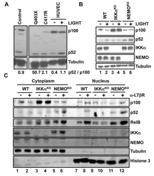
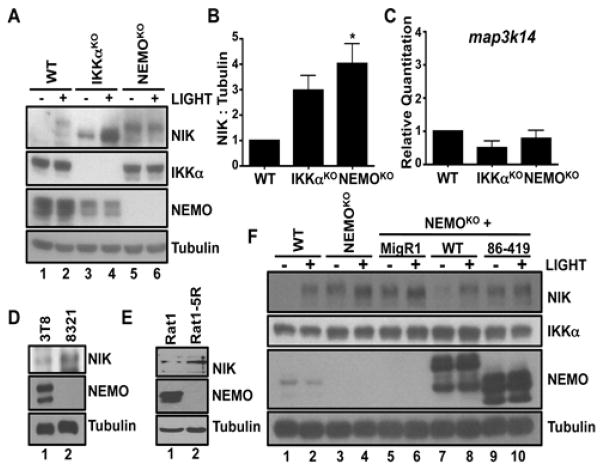
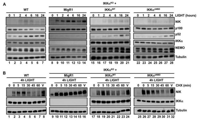
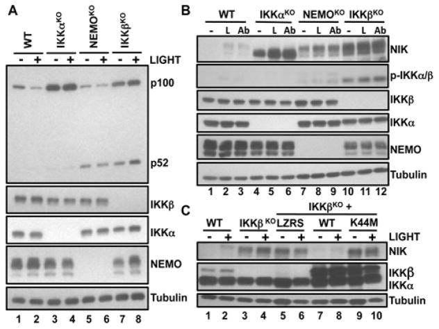
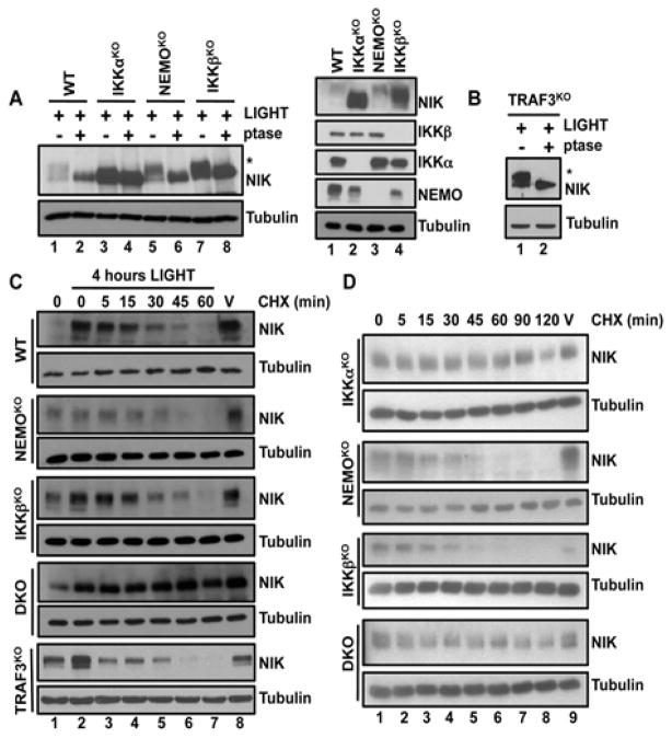
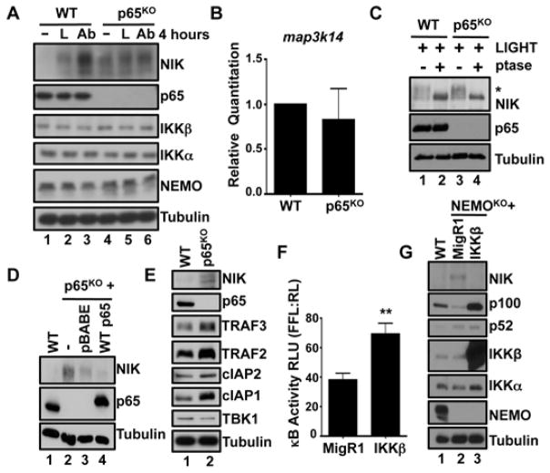
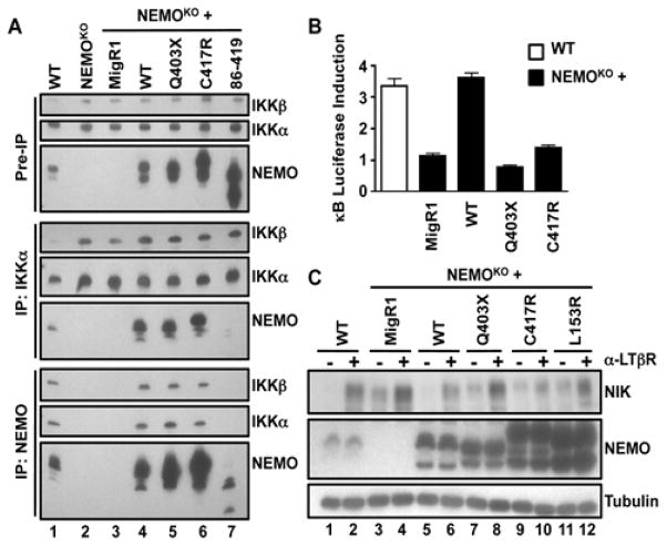
Precise regulation of nuclear factor κB (NF-κB) signaling is crucial for normal immune responses, and defective NF-κB activity underlies a range of immunodeficiencies. NF-κB is activated through two signaling cascades: the classical and noncanonical pathways. The classical pathway requires inhibitor of κB kinase β (IKKβ) and NF-κB essential modulator (NEMO), and hypomorphic mutations in the gene encoding NEMO (ikbkg) lead to inherited immunodeficiencies, collectively termed NEMO-ID. Noncanonical NF-κB activation requires NF-κB-inducing kinase (NIK) and IKKα, but not NEMO. We found that noncanonical NF-κB was basally active in peripheral blood mononuclear cells from NEMO-ID patients and that noncanonical NF-κB signaling was similarly enhanced in cell lines lacking functional NEMO. NIK, which normally undergoes constitutive degradation, was aberrantly present in resting NEMO-deficient cells, and regulation of its abundance was rescued by reconstitution with full-length NEMO, but not a mutant NEMO protein unable to physically associate with IKKα or IKKβ. Binding of NEMO to IKKα was not required for ligand-dependent stabilization of NIK or noncanonical NF-κB signaling. Rather, an intact and functional IKK complex was essential to suppress basal NIK activity in unstimulated cells. Despite interacting with IKKα and IKKβ to form an IKK complex, NEMO mutants associated with immunodeficiency failed to rescue classical NF-κB signaling or reverse the accumulation of NIK. Together, these findings identify a crucial role for classical NF-κB activity in the suppression of basal noncanonical NF-κB signaling.


Conflict of interest statement

Competing interests: The authors declare that they have no competing interests.

Figures

Fig. 1
Fig. 1. The extent of p100 processing is enhanced in NEMO-ID cells and cells lacking NEMO
(A) Lysates of PBMCs from patients with distinct NEMO mutations (Q403X and C417R), PBMCs from a healthy donor (Control) and HUVECs that were either untreated (−) or were stimulated with LIGHT for 8 hours (+) were analyzed by Western blotting with antibodies against the indicated proteins. Values reported here (in numbers beneath the blots) for the quantification of the ratio of the intensity of the p52 bands to the intensity of the p100 bands are arbitary units relative to the intensity of the tubulin bands. (B) WT, IKKαKO, and NEMOKO MEFs were either untreated (−) or were stimulated with LIGHT for 12 hours (+). Cell lysates were then analyzed by Western blotting with antibodies against the indicated proteins. (C) WT, IKKαKO, and NEMOKO MEFs were either left untreated (−) or were incubated with anti-LTβR antibody (α-LTβR) for 12 hours (+). Cytoplasmic and nuclear extracts were then prepared and analyzed by Western blotting with antibodies against the indicated proteins. Western blots are representative of three independent experiments.
Fig. 2
Fig. 2. NEMO suppresses the accumulation of NIK protein
(A) WT, IKKαKO, and NEMOKO MEFs were either left untreated (−) or incubated with LIGHT for 12 hours (+). Cell lysates were then analyzed by Western blotting with antibodies against the indicated proteins. (B) Quantification of the abundance of NIK protein relative to that of tubulin in the experiments represented by the Western blot in (A). Data are means ± SEM from three independent experiments. *P < 0.05 by one-way ANOVA and Dunnett’s post-test. (C) Analysis of the abundance of map3k14 mRNA (which encodes NIK) in WT, IKKαKO, and NEMOKO MEFs. Data are means ± SEM from four independent experiments. (D and E) Lysates from (D) WT and NEMO-deficient Jurkat cells and (E) Rat1R fibroblasts were analyzed by Western blotting with antibodies against the indicated proteins. (F) WT, NEMOKO, and NEMOKO MEFs retrovirally transduced with either control MigR1 vector or with MigR1 encoding WT NEMO or a truncated mutant NEMO (86–419) were either untreated (−) or incubated with LIGHT for 12 hours (+) before cell lysates were analyzed by Western blotting with antibodies against the indicated proteins. Western blots are representative of four independent experiments.
Fig. 3
Fig. 3. An association between NEMO and IKKα is dispensable to regulate non-canonical NF-κB
(A) WT and IKKα-deficient (IKKαKO) MEFs retrovirally transduced with either MigR1 alone or MigR1 expressing IKKαWT or IKKαΔNBD were incubated with LIGHT for the indicated times. Cell lysates were then analyzed by Western blotting with antibodies against the indicated proteins. (B) WT, IKKαKO, and IKKαKO MEFs transduced with the indicated constructs were incubated for 4 hours with LIGHT and then were treated either with cycloheximide (CHX) for the indicated times or with ethanol for 60 min as a vehicle control (V). Lysates were then analyzed by Western blotting with antibodies against the indicated proteins. Western blots are representative of four independent experiments.
Fig. 4
Fig. 4. The catalytically active IKK complex is required to suppress accumulation of NIK
(A) WT, IKKαKO, NEMOKO, and IKKβKO MEFs were either untreated (−) or incubated with LIGHT for 12 hours (+) before cell lysates were analyzed by Western blotting with antibodies against the indicated proteins. (B) The indicated MEF cell lines were either untreated (−) or were stimulated with LIGHT (L) or anti-LTβR antibody (Ab) for 12 hours before cell lysates were analyzed by Western blotting with antibodies against the indicated proteins. Active IKK was detected with an antibody specific for phosphorylated IKKα/β (p-IKKα/β). (C) WT, IKKβKO, and IKKβKO MEFs retrovirally transduced with LZRS or LZRS expressing either IKKβWT or a catalytically inactive mutant IKKβ (IKKβK44M) were either untreated (−) or incubated with LIGHT for 12 hours (+) before cell lysates were analyzed by Western blotting with antibodies against the indicated proteins. Western blots are representative of four independent experiments.
Fig. 5
Fig. 5. The IKK complex does not mediate the IKKα-dependent turnover of active NIK protein
(A) Left: The indicated cells were incubated with LIGHT for 4 hours, and then cell lysates were prepared and either left untreated (−) or incubated with λ-phosphatase (ptase; +) before being analyzed by Western blotting with antibodies against the indicated proteins. The asterisk indicates the band corresponding to phosphorylated NIK. Right: Loading controls to show the abundances of NIK, IKKα, IKKβ, and NEMO in lysates of the indicated LIGHT-stimulated cells before treatment with phosphatase. (B) TRAF3KO MEFs were processed and analyzed as described in (A). (C) WT, NEMOKO, IKKβKO, DKO, and TRAF3KO MEFs were incubated for 4 hours with LIGHT and then were either treated with cycloheximide (CHX) for the indicated times or were treated for 60 min with ethanol as a vehicle control (V). Cell lysates were analyzed by Western blotting with antibodies against the indicated proteins. (D) Unstimulated IKKαKO, NEMOKO, IKKβKO, and DKO MEFs were incubated with CHX for the indicated times or with ethanol (V) for 120 min before cell lysates were prepared and analyzed by Western blotting with antibodies against the indicated proteins. Western blots are representative of four independent experiments.
Fig. 6
Fig. 6. Classical NF-κB–dependent gene expression is required to regulate basal NIK abundance
(A) WT or p65KO MEFs were left unstimulated or were treated with either LIGHT (L) or the anti-LTβR antibody (Ab) for 4 hours. Cell lysates were then analyzed by Western blotting with antibodies against the indicated proteins. (B) RNA from WT or p65KO MEFs was isolated and analyzed by quantitative RT-PCR to determine the abundance of map3k14 mRNA in comparison to that of actb mRNA (which encodes β-actin). Data are means ± SEM from three independent experiments. (C) WT and p65KO MEFs were stimulated with LIGHT for 4 hours. Whole-cell extracts were then left untreated or were treated with λ phosphatase and analyzed by Western blotting with antibodies against the indicated proteins. The asterisk indicates the band corresponding to phosphorylated NIK. (D) p65KO MEFs were transduced with empty pBABE vector or the pBABE vector encoding WT p65. Cell lysates were then analyzed by Western blotting with antibodies against the indicated proteins. (E) Whole-cell extracts from WT or p65KO MEFs were analyzed by Western blotting with antibodies against the indicated proteins. (F) NEMOKO MEFs stably transduced with either MigR1 or MigR1 encoding WT IKKβ were transfected with the pBIIx-κB firefly luciferase (FFL) and the TK renilla luciferase (RL) vectors. Twenty-four hours later, classical NF-κB transcriptional activity was determined as the fluorescence ratio of FFL:RL. Data are means ± SEM of six replicates from two independent experiments. **P < 0.01 by student’s two-tailed t-test. (G) WT or NEMOKO MEFs stably transduced with either MigR1 or MigR1 encoding WT IKKβ were analyzed by Western blotting with antibodies against the indicated proteins. Western blots are representative of four independent experiments.
Fig. 7
Fig. 7. Noncanonical NF-κB signaling is enhanced in cells containing hypomorphic ikbkg mutations
(A) WT, NEMOKO, and NEMOKO MEFs retrovirally transduced with MigR1 or MigR1 encoding WT NEMO or the indicated NEMO mutants were lysed and then subjected to immunoprecipitation (IP) with anti-IKKα and anti-NEMO antibodies. Cell lysates and the immunoprecipitated samples were analyzed by Western blotting with antibodies against the indicated proteins. (B) WT and NEMOKO cells were transfected with the pBIIx-κB firefly luciferase (FFL) and the TK Renilla luciferase (RL) vectors. Twenty-four hours later, cells were either left untreated or were treated with TNF-α for 5 hours. NF-κB transcriptional activity was determined by measuring the fluorescence ratio of FFL:RL, and the extent of κB luciferase induction was defined as the fold-difference between the TNF-α–stimulated samples and the controls. Data are means ± SD of four replicates from one of three independent experiments. (C) WT and NEMOKO MEFs retrovirally transduced with MigR1 or MigR1 encoding WT NEMO or the indicated NEMO mutants were either untreated (−) or incubated with anti-LTβR antibody for 8 hours (+) before being lysed and analyzed by Western blotting with antibodies against the indicated proteins. Western blots are representative of four independent experiments.

Similar articles

Cited by

References

    1. Hayden MS, Ghosh S. Shared principles in NF-kappaB signaling. Cell. 2008;132:344–362. published online EpubFeb 8. - PubMed
    1. Hayden MS, Ghosh S. NF-kappaB in immunobiology. Cell Res. 2011;21:223–244. published online EpubFeb. - PMC - PubMed
    1. Kawai T, Nishikomori R, Heike T. Diagnosis and treatment in anhidrotic ectodermal dysplasia with immunodeficiency. Allergology international: official journal of the Japanese Society of Allergology. 2012;61:207–217. - PubMed
    1. Solt LA, May MJ. The IkappaB kinase complex: master regulator of NF-kappaB signaling. Immunol Res. 2008;42:3–18. - PMC - PubMed
    1. Rudolph D, Yeh WC, Wakeham A, Rudolph B, Nallainathan D, Potter J, Elia AJ, Mak TW. Severe liver degeneration and lack of NF-kappaB activation in NEMO/IKKgamma-deficient mice. Genes Dev. 2000;14:854–862. - PMC - PubMed

Publication types

MeSH terms

Substances

LinkOut - more resources