. 2014 May 2;289(18):12300-12.
doi: 10.1074/jbc.M113.529164. Epub 2014 Feb 4.

Fluoroquinolone-gyrase-DNA complexes: two modes of drug binding

Affiliations

Fluoroquinolone-gyrase-DNA complexes: two modes of drug binding

Arkady Mustaev et al. J Biol Chem. .

Abstract

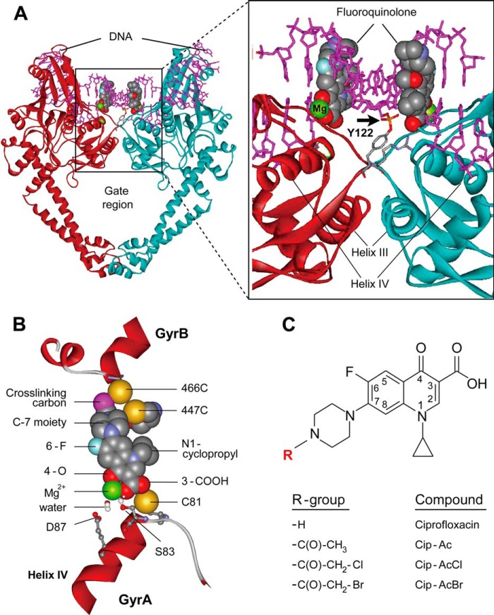
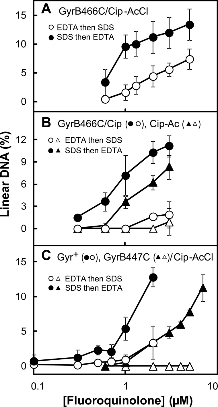
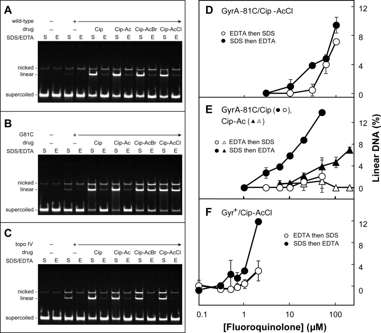
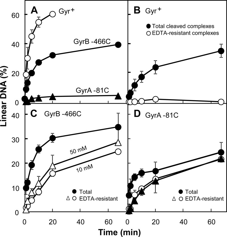
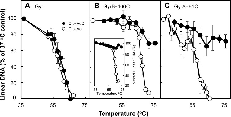
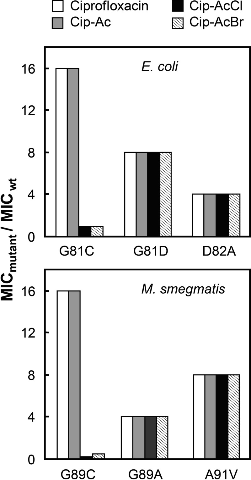
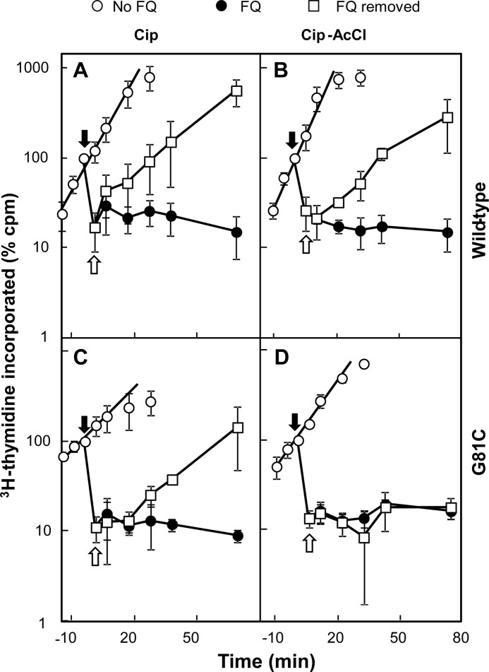
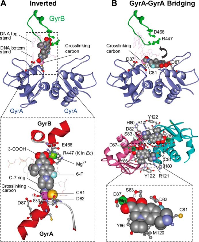
DNA gyrase and topoisomerase IV control bacterial DNA topology by breaking DNA, passing duplex DNA through the break, and then resealing the break. This process is subject to reversible corruption by fluoroquinolones, antibacterials that form drug-enzyme-DNA complexes in which the DNA is broken. The complexes, called cleaved complexes because of the presence of DNA breaks, have been crystallized and found to have the fluoroquinolone C-7 ring system facing the GyrB/ParE subunits. As expected from x-ray crystallography, a thiol-reactive, C-7-modified chloroacetyl derivative of ciprofloxacin (Cip-AcCl) formed cross-linked cleaved complexes with mutant GyrB-Cys(466) gyrase as evidenced by resistance to reversal by both EDTA and thermal treatments. Surprisingly, cross-linking was also readily seen with complexes formed by mutant GyrA-G81C gyrase, thereby revealing a novel drug-gyrase interaction not observed in crystal structures. The cross-link between fluoroquinolone and GyrA-G81C gyrase correlated with exceptional bacteriostatic activity for Cip-AcCl with a quinolone-resistant GyrA-G81C variant of Escherichia coli and its Mycobacterium smegmatis equivalent (GyrA-G89C). Cip-AcCl-mediated, irreversible inhibition of DNA replication provided further evidence for a GyrA-drug cross-link. Collectively these data establish the existence of interactions between the fluoroquinolone C-7 ring and both GyrA and GyrB. Because the GyrA-Gly(81) and GyrB-Glu(466) residues are far apart (17 Å) in the crystal structure of cleaved complexes, two modes of quinolone binding must exist. The presence of two binding modes raises the possibility that multiple quinolone-enzyme-DNA complexes can form, a discovery that opens new avenues for exploring and exploiting relationships between drug structure and activity with type II DNA topoisomerases.

Keywords: Antibiotic Action; Bacteria; Bacterium; Cleaved Complexes; Cross-linking; DNA Topoisomerase; Enzyme Inhibitors; Escherichia coli; Mycobacterium smegmatis; Resistance Mutations.


Figures

FIGURE 1.
FIGURE 1.
Structures of type II topoisomerase-DNA-quinolone complex and quinolones. A, cleaved complex. The side view of the three-dimensional structure of Acinetobacter baumannii topoisomerase IV bound to DNA and moxifloxacin (Protein Data Bank code 2XKK) is shown in which the DNA gate region is illustrated in an expanded view (right). Moxifloxacin is depicted in a space-filling representation; the arrow indicates the covalent bond between the DNA end and GyrA-Tyr122. One GyrA subunit is shown in maroon, the other is shown in turquoise, and DNA is pink. GyrB is omitted for clarity. B, enlargement of the quinolone-binding region. The proximity of GyrB cysteine substitutions to the cross-linking carbon of Cip-AcCl is shown. Other fluoroquinolone moieties and GyrA amino acids are present to provide orientation. C, structures of quinolones. Ciprofloxacin is shown along with the modified derivatives used in this work.
FIGURE 2.
FIGURE 2.
Evidence for cross-linking of GyrB-E466C gyrase to Cip-AcCl. Mixtures containing gyrase, gel-purified supercoiled pBR322 DNA, and ciprofloxacin-based derivatives were incubated at 37 °C. Then reaction mixtures were treated with either SDS and proteinase K (filled symbols) to release DNA breaks generated by drug-gyrase action or with EDTA (empty symbols) to reseal DNA breaks. The SDS-treated samples were then treated with EDTA, and the EDTA samples were treated with SDS and proteinase K before samples were subjected to gel electrophoresis to separate the DNA species. The percentage of linear DNA within each sample was determined as a measure of DNA breaks generated by gyrase-drug action at the indicated drug concentrations. In the data shown, concentrations of gyrase and DNA were 20 and 7.4 nm, respectively; reactions were incubated for 15 min. Error bars indicate S.D. A shows GyrB-E466C gyrase incubated with the cross-linking agent Cip-AcCl; B shows results for GyrB-E466C gyrase with two non-cross-linking agents, ciprofloxacin (circles) and Cip-Ac (triangles); and C shows wild-type gyrase (circles) or GyrB-Cys447 gyrase (triangles) with the cross-linking agent Cip-AcCl. Examination of a variety of enzyme:DNA ratios and incubation times produced results similar to those shown. The number of total experiments and those used for the data shown (chosen so panels had the same conditions), respectively, were 14 and 5 for the comparison of A, 7 and 4 for ciprofloxacin in B, 4 and 4 for Cip-Ac in B, 3 and 3 for GyrB-Cys447 gyrase in C, and 11 and 4 for wild-type gyrase in C.
FIGURE 3.
FIGURE 3.
Evidence for cross-linking of GyrA-G81C gyrase to haloacetyl derivatives of ciprofloxacin. Reaction mixtures containing gyrase or topoisomerase IV, supercoiled pBR322 DNA, and derivatives of ciprofloxacin were incubated at 37 °C as described under “Experimental Procedures.” A–C, display of plasmid DNA species following incubation with derivatives of ciprofloxacin and purified gyrase or topoisomerase IV. Concentrations of enzymes and drugs were chosen to obtain similar amounts of linear DNA. The DNA cleavage assay used 5 nm plasmid pBR322 DNA and 7.5 nm wild-type gyrase (A), 25 nm GyrA-G81C gyrase (B), or 7.5 nm topoisomerase IV (C). Ciprofloxacin was used at 1 μm for wild-type gyrase; it was 25 μm for GyrA-G81C gyrase and topoisomerase IV. Cip-Ac, Cip-AcBr, and Cip-AcCl were used at 5 μm for wild-type gyrase and 125 μm for GyrA-G81C gyrase and topoisomerase IV. S and E indicate DNA products from reaction mixtures without and with 50 mm EDTA-mediated reversal, respectively. The assay was repeated twice with identical results (less than 2% difference in the percentage of linear DNA). The effect of drug concentration on the recovery of linear DNA is shown in D–F; data were obtained as in Fig. 2 except that GyrA-G81C was substituted for GyrB-E466C. For the data shown, concentrations of enzyme and DNA were 20 and 7.4 nm, respectively; reactions were incubated for 30 min at 37 °C and then treated either with 50 mm EDTA and then SDS (empty symbols) or with SDS first and then EDTA (filled symbols). D shows the effect of the cross-linking agent Cip-AcCl with GyrA-G81C gyrase, E shows results for non-cross-linking ciprofloxacin with GyrA-G81C gyrase (circles) and for non-cross-linking Cip-Ac with GyrA-G81C gyrase (triangles), and F shows results for wild-type gyrase with the cross-linking agent Cip-AcCl. Examination of a variety of enzyme:DNA ratios and incubation times produced results similar to those shown. The number of total experiments and those used for the data shown (chosen so panels had the same incubation conditions), respectively, were 9 and 4 for the comparison of D, 10 and 2 for ciprofloxacin in E, 8 and 4 for Cip-Ac in E, and 7 and 2 for wild-type gyrase and the cross-linking agent Cip-AcCl in F. Total DNA recovery was unaffected by drug concentration, indicating that loss of specific bands was not due to preferential recovery. Error bars indicate S.D.
FIGURE 4.
FIGURE 4.
Time course of formation of cleaved complexes containing Cip-AcCl. Reaction mixtures were prepared as in Fig. 2 using wild-type, GyrA-G81C, or GyrB-E466C gyrase, pBR322, and the cross-linking compound Cip-AcCl. Mixtures were then incubated at 37 °C for various times and processed to reveal the percentage of total linear DNA or the percentage of linear DNA remaining after reversal by EDTA. A, total cleaved complex formation. Gyrase (40 nm), supercoiled pBR322 DNA (3.7 nm), and Cip-AcCl (10 μm) were incubated for the indicated times followed by treatment with SDS and proteinase K for an additional 15 min. Empty circles, wild-type gyrase; filled circles, GyrB-E466C gyrase; filled triangles, GyrA-G81C gyrase. B, EDTA-resistant and total complexes with wild-type gyrase. Reaction mixtures, prepared as in A but with 2 μm Cip-AcCl, were incubated for the indicated times followed by an additional incubation with 10 mm EDTA for 15 min and then with SDS-proteinase K for another 15 min (empty circles). Parallel samples of reaction mixtures were incubated with SDS-proteinase before EDTA treatment (filled circles) as a measure of total complexes. C, EDTA-resistant and total complexes with GyrB-E466C gyrase. Reaction mixtures with GyrB-E466C gyrase, prepared as in A, were incubated for the indicated times followed by an additional incubation with 10 mm EDTA (empty circles) or 50 mm EDTA (empty triangles) for 15 min and then with SDS-proteinase K for another 15 min (filled circles). Parallel samples of reaction mixtures were incubated with SDS-proteinase before EDTA treatment (filled circles) to assess total complexes. D, EDTA-resistant and total complexes with GyrA-G81C gyrase. Conditions and treatments were as in C except for the use of 40 μm Cip-AcCl and 80 nm GyrA-G81C gyrase. Empty circles, 10 mm EDTA before SDS; filled triangles, 50 mm EDTA before SDS; filled circles, SDS before EDTA. Error bars indicate S.D. for four experiments.
FIGURE 5.
FIGURE 5.
Effect of temperature on DNA resealing. Reaction mixtures containing gyrase, plasmid DNA, and derivatives of ciprofloxacin were incubated for 45 min at 37 °C followed by an additional 5-min incubation at the indicated temperature. Reactions were stopped by chilling on ice; additional SDS-proteinase K treatment and electrophoretic analysis were as described under “Experimental Procedures.” The percentage of DNA in the linear form was determined for each temperature; data were then normalized to the percentage of linear DNA observed at 37 °C to allow comparison of thermal resealing curves containing different levels of linear DNA prior to thermal treatment. A, wild-type gyrase (40 nm) and pBR322 (3.7 nm) were incubated with Cip-Ac (6 μm; empty circles) or Cip-AcCl (6 μm; filled circles). B, GyrB-E466C gyrase (40 nm) and pBR322 DNA (3.7 nm) were incubated with Cip-Ac (80 μm; empty circles) or Cip-AcCl (10 μm; filled circles). The inset shows the effect of thermal incubation on the sum of linear plus nicked DNA for complexes formed with Cip-Ac (empty circles) and Cip-AcCl (filled circles). C, GyrA-G81C gyrase (80 nm) and pBR322 (3.7 nm) were incubated with Cip-Ac (700 μm; empty circles) or Cip-AcCl (500 μm; filled circles). Elevated quinolone concentrations were required to obtain linear DNA at levels similar to those used in A and B (above 30%). Error bars indicate S.D. for the following number of determinations: A, Cip-AcCl, 4; Cip-Ac, 3; B, Cip-AcCl, 4; Cip-Ac, 3; C, Cip-AcCl, 8; Cip-Ac, 5.
FIGURE 6.
FIGURE 6.
Effect of chloroacetyl and bromoacetyl substituents attached to ciprofloxacin on growth inhibition of GyrA Cys variants. The MIC was determined with the indicated mutants for ciprofloxacin derivatives (white bars, ciprofloxacin; gray bars, Cip-Ac; black bars, Cip-AcCl; striped bars, Cip-AcBr) with E. coli (upper panel) and M. smegmatis (lower panel). Data are expressed as multiples of values for MIC determined with wild-type E. coli strain DM4100 (MIC = 0.01 μg/ml for ciprofloxacin, 0.3 μg/ml for Cip-Ac, 0.25 μg/ml for Cip-AcCl, and 0.5 μg/ml for Cip-AcBr) or wild-type M. smegmatis strain mc2155 (0.1 μg/ml for ciprofloxacin, 0.16 μg/ml for Cip-Ac, 0.5 μg/ml for Cip-AcCl, and 0.5 μg/ml for Cip-AcBr). For strain numbers, see Table 1. Three independent experiments gave identical results, precluding determination of error bars.
FIGURE 7.
FIGURE 7.
Cip-AcCl preferentially blocks reversal of DNA synthesis inhibition with GyrA81 variant. E. coli strains KD2372 (gyrA+) and KD3052 (GyrA-G81C) were grown as liquid cultures that were treated at time 0 (filled arrow) with either ciprofloxacin (A and C) or Cip-AcCl (B and D) at 10× MIC. Five min later (empty arrow) an aliquot of treated cells was collected by filtration, and the cells were quickly resuspended in drug-free medium. At various times, aliquots were removed, and the DNA synthesis rate was measured as described under “Experimental Procedures.” Empty circles, untreated control; filled circles, treated with fluoroquinolone (FQ) at time 0; empty squares, treated with fluoroquinolone for 5 min, and then drug was removed by filtration. Error bars indicate S.D. from four determinations.
FIGURE 8.
FIGURE 8.
Models for binding of ciprofloxacin to gyrase-DNA complexes with interaction between fluoroquinolone C-7 ring and GyrA81. A, inverted configuration of the published x-ray crystal structure (Protein Data Bank code 2XKK). In the enlargement, GyrB447 is Arg in M. smegmatis and Lys in E. coli. B, GyrA-GyrA bridging model (from Protein Data Bank code 1AB4). The top portion shows one molecule of cross-linked ciprofloxacin in a bridging pocket. The curved arrow indicates the rotation needed to achieve the inverted structure shown in A. The center portion shows two antiparallel pockets filled with fluoroquinolone. The bottom portion shows a detail of nearby GyrA amino acid residues; GyrA87 and GyrA81 are on different GyrA subunits. In both panels, the cross-linking carbon is labeled.

Similar articles

Cited by

References

    1. Nitiss J. L. (2009) Targeting DNA topoisomerase II in cancer chemotherapy. Nat. Rev. Cancer 9, 338–350 - PMC - PubMed
    1. Champoux J. J. (2001) DNA topoisomerases: structure, function, and mechanism. Annu. Rev. Biochem. 70, 369–413 - PubMed
    1. Wang J. C. (2002) Cellular roles of DNA topoisomerases: a molecular perspective. Nat. Rev. Mol. Cell Biol. 3, 430–440 - PubMed
    1. Mizuuchi K., Fisher L. M., O'Dea M. H., Gellert M. (1980) DNA gyrase action involves the introduction of transient double-strand breaks into DNA. Proc. Natl. Acad. Sci. U.S.A. 77, 1847–1851 - PMC - PubMed
    1. Kreuzer K. N., Cozzarelli N. R. (1980) Formation and resolution of DNA catenanes by DNA gyrase. Cell 20, 245–254 - PubMed

Publication types

MeSH terms