

Time-of-flight magnetic resonance angiography for follow-up of coil embolization with enterprise stent for intracranial aneurysm: usefulness of source images
- PMID: 24497808
- PMCID: PMC3909851
- DOI: 10.3348/kjr.2014.15.1.161
Time-of-flight magnetic resonance angiography for follow-up of coil embolization with enterprise stent for intracranial aneurysm: usefulness of source images
Abstract
Objective: The aim of this study was to determine the interobserver and intermodality agreement in the interpretation of time-of-flight (TOF) MR angiography (MRA) for the follow-up of coiled intracranial aneurysms with the Enterprise stent.
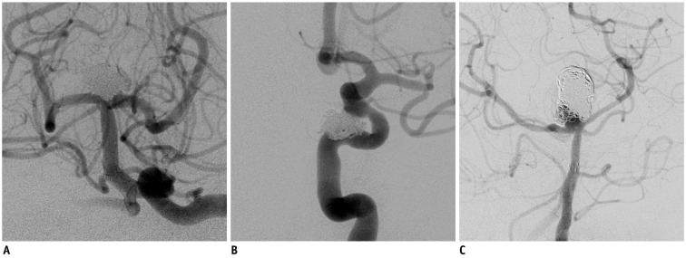
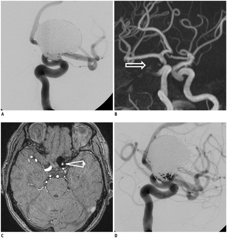
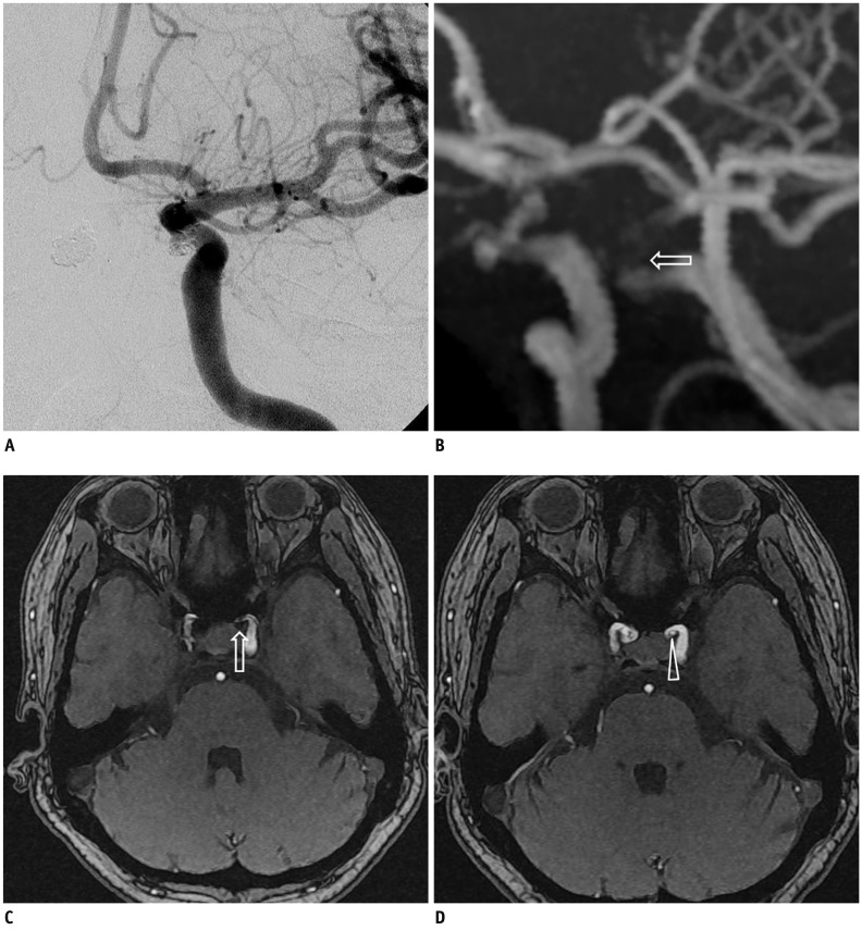
Materials and methods: Two experienced neurointerventionists independently reviewed the follow-up MRA studies of 40 consecutive patients with 44 coiled aneurysms. All aneurysms were treated with assistance from the Enterprise stent and the radiologic follow-up intervals were greater than 6 months after the endovascular therapy. Digital subtraction angiography (DSA) served as the reference standard. The degree of aneurysm occlusion was determined by an evaluation of the maximal intensity projection (MIP) and source images (SI) of the TOF MRA. The capability of the TOF MRA to depict the residual flow within the coiled aneurysms and the stented parent arteries was compared with that of the DSA.
Results: DSA showed stable occlusions in 25 aneurysms, minor recanalization in 8, and major recanalization in 11. Comparisons between the TOF MRA and conventional angiography showed that the MIP plus SI had almost perfect agreement (κ = 0.892, range 0.767 to 1.000) and had better agreement than with the MIP images only (κ = 0.598, range 0.370 to 0.826). In-stent stenosis of more than 33% was observed in 5 cases. Both MIP and SI of the MRA showed poor depiction of in-stent stenosis compared with the DSA.
Conclusion: TOF MRA seemed to be reliable in screening for aneurysm recurrence after coil embolization with Enterprise stent assistance, especially in the evaluation of the SI, in addition to MIP images in the TOF MRA.
Keywords: Aneurysm; Coiling; Embolization; MRA; Stent.
Figures
References
-
- Molyneux A, Kerr R, Stratton I, Sandercock P, Clarke M, Shrimpton J, et al. International Subarachnoid Aneurysm Trial (ISAT) of neurosurgical clipping versus endovascular coiling in 2143 patients with ruptured intracranial aneurysms: a randomised trial. Lancet. 2002;360:1267–1274. - PubMed
-
- Lavoie P, Gariépy JL, Milot G, Jodoin S, Bédard F, Trottier F, et al. Residual flow after cerebral aneurysm coil occlusion: diagnostic accuracy of MR angiography. Stroke. 2012;43:740–746. - PubMed
-
- Piotin M, Blanc R, Spelle L, Mounayer C, Piantino R, Schmidt PJ, et al. Stent-assisted coiling of intracranial aneurysms: clinical and angiographic results in 216 consecutive aneurysms. Stroke. 2010;41:110–115. - PubMed
-
- Takayama K, Taoka T, Nakagawa H, Myouchin K, Wada T, Sakamoto M, et al. Usefulness of contrast-enhanced magnetic resonance angiography for follow-up of coil embolization with the enterprise stent for cerebral aneurysms. J Comput Assist Tomogr. 2011;35:568–572. - PubMed
MeSH terms
LinkOut - more resources
Full Text Sources
Other Literature Sources
Medical
