. 2014 Jan 30;10(1):e1004139.
doi: 10.1371/journal.pgen.1004139. eCollection 2014 Jan.

Large inverted duplications in the human genome form via a fold-back mechanism

Affiliations

Large inverted duplications in the human genome form via a fold-back mechanism

Karen E Hermetz et al. PLoS Genet. .

Abstract

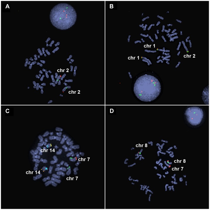
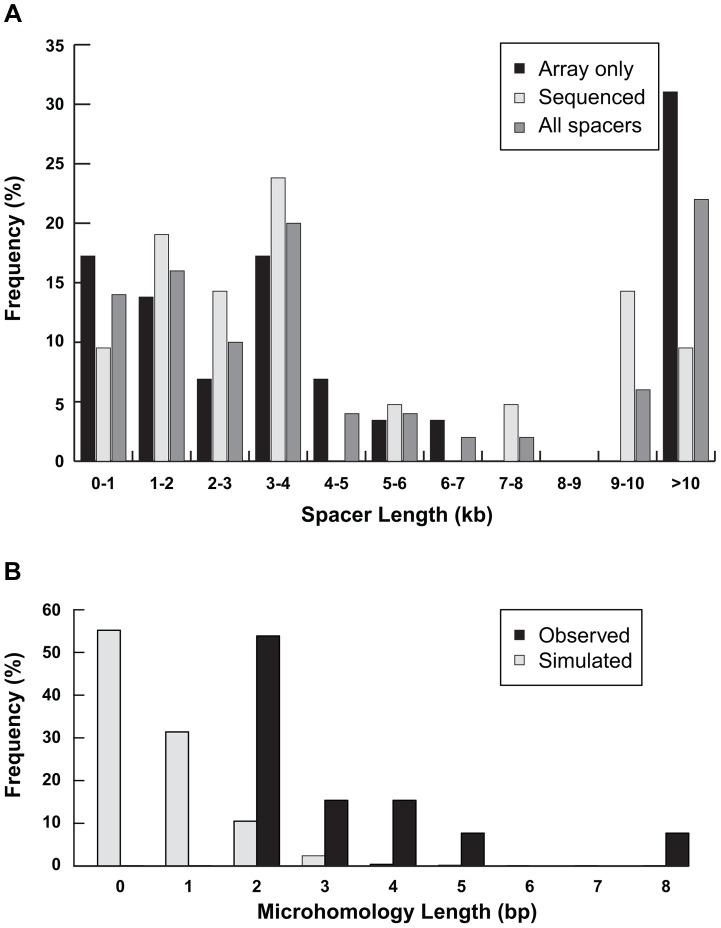
Inverted duplications are a common type of copy number variation (CNV) in germline and somatic genomes. Large duplications that include many genes can lead to both neurodevelopmental phenotypes in children and gene amplifications in tumors. There are several models for inverted duplication formation, most of which include a dicentric chromosome intermediate followed by breakage-fusion-bridge (BFB) cycles, but the mechanisms that give rise to the inverted dicentric chromosome in most inverted duplications remain unknown. Here we have combined high-resolution array CGH, custom sequence capture, next-generation sequencing, and long-range PCR to analyze the breakpoints of 50 nonrecurrent inverted duplications in patients with intellectual disability, autism, and congenital anomalies. For half of the rearrangements in our study, we sequenced at least one breakpoint junction. Sequence analysis of breakpoint junctions reveals a normal-copy disomic spacer between inverted and non-inverted copies of the duplication. Further, short inverted sequences are present at the boundary of the disomic spacer and the inverted duplication. These data support a mechanism of inverted duplication formation whereby a chromosome with a double-strand break intrastrand pairs with itself to form a "fold-back" intermediate that, after DNA replication, produces a dicentric inverted chromosome with a disomic spacer corresponding to the site of the fold-back loop. This process can lead to inverted duplications adjacent to terminal deletions, inverted duplications juxtaposed to translocations, and inverted duplication ring chromosomes.


Conflict of interest statement

The authors have declared that no competing interests exist.

Figures

Figure 1
Figure 1. Inverted duplication organization.
(A) Model of duplicated sequences (orange arrows) separated by disomic spacer sequence (grey line). The end of the inverted duplication may terminate in a telomere (black triangle) or a translocated chromosome (blue). The site of the terminal deletion is shown relative to a normal chromosome. (B) EGL044's inverted duplication of chromosome 2 is detectable by chromosome banding. (C) The 5.8-Mb terminal deletion and 42-Mb inverted duplication of chromosome 2 are detectable by low-resolution array CGH . Note that the 2,047-bp spacer region is not visible. Log2 ratios of oligonucleotide probes are indicated by dots; normal-copy number (black), duplication (red), and deletion (green) regions are shown. (D) PCR of the disomy-inversion junction (lane 2) and the inversion-telomere junction (lane 4) amplifies genomic DNA from EGL044, but not control genomic DNA (lanes 3 and 5). Lane 1 is GeneRuler 1 kb Plus DNA ladder (Thermo Scientific Fermentas #SM1333).
Figure 2
Figure 2. FISH analysis of inverted duplication translocation chromosomes.
(A) EGL398's 3.1-Mb duplication of 2q37 is visible by interphase FISH. BAC probes RP11-206J15 (red) and RP11-1415N13 (green) hybridize to the duplicated and control regions on chromosome 2, respectively. Three red signals in the interphase nucleus indicate a duplication of chromosome 2q37. (B) BAC probes RP11-798H13 (red) and RP11-380E2 (green) hybridize to the ends of the normal chromosomes 1p and the end of the inverted duplication translocation chromosome in EGL398. (C) EGL399's terminal deletion of 7q is detected as loss of a red signal. Vysis ToTelVysion mix 7 (Abbott Molecular, #05J05-001) probes hybridize to the ends of chromosomes 7p (green), 7q (red), and 14q (yellow). The blue signals correspond to a control probe that hybridizes to chromosome 14q11. (D) BAC RP11-341D4 (red) hybridizes to the normal chromosomes 8p and the translocation of 8p on EGL399's inverted duplication translocation between chromosomes 7 and 8. The green signal corresponds to alpha satellite from the centromere of chromosome 8.
Figure 3
Figure 3. High-resolution array CGH identifies spacers.
5-Mb (left) and 400-kb (right) views of high-resolution array CGH data from (A) 18q-6c, (B) EGL106, (C) EGL104, (D) 18q-233c, and (E) SG_Tel_010 show 1,866-bp, 3,138-bp, 14,779-bp, 70,466-bp, and 14,779-bp spacers, respectively. Log2 ratios of probe signal intensity are shown as black dots. Boxed region on the left is expanded on the right. Red arrows point out disomic spacer regions between deleted and duplicated segments. Spacer sizes were determined by sequencing breakpoint junctions in (A)–(D), whereas the spacer in (E) was sized using breakpoints determined by array CGH only.
Figure 4
Figure 4. Inverted duplication junctions.
(A) Location of disomy-inversion and inversion-telomere junctions in an inverted duplication terminal deletion chromosome. (B) 18q-233c's disomy-inversion junction spans a hybrid LINE made up of L1PA2 and L1Hs elements. On a normal chromosome 18, these elements are positioned in opposite orientation. (C) Local genomic context of 18q-233c's spacer and breakpoints relative to the reference genome assembly. The distal end of the disomic spacer (grey box) includes the L1PA2, and the proximal region corresponding to the beginning of the inverted duplication (orange box) includes the L1Hs. The disomy-inversion junction sequence (black rectangles with white arrows) aligns to the distal end of the spacer (positions 1–465 of the junction) and the start of the inverted duplication (positions 140–834 of the junction). Interspersed repeats are shown as black rectangles. No segmental duplications are present in the breakpoint regions.
Figure 5
Figure 5. Characterization of spacers.
(A) Distribution of lengths of spacers measured by high-resolution array CGH only (n = 29) or junction sequencing (n = 21) are plotted separately. The distribution of all 50 spacer lengths is also shown. (B) The amount of inverted microhomology observed at 13 sequenced disomy-inverted duplication junctions—2 bp (n = 7), 3 bp (n = 2), 4 bp (n = 2), 5 bp (n = 1), or 8 bp (n = 1)—are shown relative to the microhomology detected for 1,000 simulated spacers (see Methods).
Figure 6
Figure 6. Complex junctions from EGL106 and 18q-65c.
Insertion orientation (+/−) is indicated relative to the reference genome. (A) Alignment of telomere (black), inverted duplication (orange), inserted sequence (blue), and junction sequence (EGL106) from the telomere-inversion junction is shown above. The inverted duplication, disomic sequence (grey), and inversion-disomy junction sequence (EGL106) alignment is shown below. Microhomology at the junction is boxed. (B) Above, disomic, inserted, and inverted duplication sequences are aligned to the disomy-inversion junction sequence (18q-65c). Below, inverted duplication and telomere sequences are aligned to the inversion-telomere junction sequence (18q-65c). Inserted sequences and their neighboring homologous sequences are underlined.
Figure 7
Figure 7. Fold-back model of inverted duplication formation.
(A) 5′ and 3′ strands of the chromosome with telomeres (triangles) and centromere (circle) are shown. Short inverted sequences (grey rectangles with arrows) lie adjacent to the terminal deletion breakpoint. The inverted duplication mechanism occurs as described in the Discussion. The resulting inverted duplication is indicated by orange arrows. (B) After a breakage-fusion-bridge cycle, the inverted duplication chromosome may be repaired as a terminal deletion, translocation, or ring chromosome.

References

    1. Weleber RG, Verma RS, Kimberling WJ, Fieger HG Jr, lubs HA (1976) Duplication-deficiency of the short arm of chromosome 8 following artificial insemination. Annales de genetique 19: 241–247. - PubMed
    1. Zuffardi O, Bonaglia M, Ciccone R, Giorda R (2009) Inverted duplications deletions: underdiagnosed rearrangements?? Clinical genetics 75: 505–513. - PubMed
    1. Rudd MK (2011) Structural variation in subtelomeres. In: Feuk L, editor. Genomic Structural Variants: Methods and Protocols. New York: Springer Science+Business Media, LLC.
    1. Ballif BC, Yu W, Shaw CA, Kashork CD, Shaffer LG (2003) Monosomy 1p36 breakpoint junctions suggest pre-meiotic breakage-fusion-bridge cycles are involved in generating terminal deletions. Hum Mol Genet 12: 2153–2165. - PubMed
    1. Bonaglia MC, Giorda R, Massagli A, Galluzzi R, Ciccone R, et al. (2009) A familial inverted duplication/deletion of 2p25.1–25.3 provides new clues on the genesis of inverted duplications. European journal of human genetics : EJHG 17: 179–186. - PMC - PubMed

Publication types

MeSH terms

Associated data