. 2014 Jan 31;9(1):e85926.
doi: 10.1371/journal.pone.0085926. eCollection 2014.

CXCR7 is highly expressed in acute lymphoblastic leukemia and potentiates CXCR4 response to CXCL12

Affiliations

CXCR7 is highly expressed in acute lymphoblastic leukemia and potentiates CXCR4 response to CXCL12

Rita de Cássia Carvalho Melo et al. PLoS One. .

Erratum in

  • PLoS One. 2014;9(3):e91365

Abstract

Recently, a novel CXCL12-binding receptor, has been identified. This CXCL12-binding receptor commonly known as CXCR7 (CXC chemokine receptor 7), has lately, based on a novel nomenclature, has received the name ACKR3 (atypical chemokine receptor 3). In this study, we aimed to investigate the expression of CXCR7 in leukemic cells, as well as its participation in CXCL12 response. Interesting, we clearly demonstrated that CXCR7 is highly expressed in acute lymphoid leukemic cells compared with myeloid or normal hematopoietic cells and that CXCR7 contributed to T-acute lymphoid leukemic cell migration induced by CXCL12. Moreover, we showed that the cellular location of CXCR7 varied among T-lymphoid cells and this finding may be related to their migration capacity. Finally, we hypothesized that CXCR7 potentiates CXCR4 response and may contribute to the maintenance of leukemia by initiating cell recruitment to bone marrow niches that were once occupied by normal hematopoietic stem cells.


Conflict of interest statement

Competing Interests: The authors have declared that no competing interests exist.

Figures

Figure 1
Figure 1. Schematic model of flow cytometric analysis.
A) An FSC/SSC gate and anti-CD45+/SSC was created around the viable lymphocyte population for further analysis of CD3+CD4+, CD3+ CD8+ subsets and CD19+ cells. B) An FSC/SSC and anti-CD45+/SSC gates were created around the viable granulocyte population for further analysis of CD14+ cells and CD16+ cells.
Figure 2
Figure 2. CXCR7 has higher expressed in ALL.
Quantitative expression of CXCR7 mRNA in patient cells relative to healthy donor cells and among different patient groups. Real time RT-PCR was performed on cDNA from the samples of patients with hematopoietic malignancies or from bone marrow samples from healthy donors. Each dot indicates the relative CXCR7 expression for each patient. Horizontal lines represent medians. mRNA expression levels of CXCR7 were normalized by HPRT and GAPDH endogenous control. A) CXCR7 mRNA was highly upregulated in BM samples from ALL patients compared to normal hematopoietic cells samples (P<0.0001) and to MDS and AML patients samples (P<0.0001). There was no significant difference in CXCR7 expression among patients with MDS, AML, and normal hematopoietic cells. B) Among ALL-diagnosed patients, CXCR7 expression was more pronounced in the T-ALL subtype; Mann-Whitney test.
Figure 3
Figure 3. CXCR7 positively correlates with the percentage of blasts in the bone marrow.
Correlation of log-transformed relative expression of CXCR7/HPRT-GAPDH and the percentage of blasts in the bone marrow of MDS, AML and ALL patients showed CXCR7 expression levels to be positively correlated with bone marrow blast counts (P = 0.004). Two-tailed Spearmans correlation. The number of individuals is shown in the figure.
Figure 4
Figure 4. Higher expression of CXCR7 in T-acute lymphoid leukemia lines MOLT4 and Jurkat.
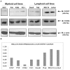
A) Western blot analysis of CXCR7 protein levels in myeloid (U937, P39, K562 and KG-1), B-lymphoid (Daudi, Raji) and T-lymphoid (MOLT4 and Jurkat) cell lines. Total cell extracts were blotted with antibodies against CXCR7 (42 kDa), CXCR4 (42 kDa) or β-actin (42 kDa), as a control for equal sample loading, and developed with the ECL Western Blot Analysis System. CXCR7 protein was detectable in all acute leukemia cell lines; however CXCR7 was more expressed in the T-acute lymphoid cell lines MOLT4 and Jurkat when compared to other cell lines. CXCR4 proteins levels were homogeneous in all cell lines analyzed. B) Quantitative expression of CXCR7 mRNA in leukemic cells lines. mRNA expression levels of CXCR7 were normalized by HPRT and GAPDH endogenous control. CXCR7 mRNA was more expressed in T-acute lymphoid cell lines MOLT4 and Jurkat when compared to other cell lines.
Figure 5
Figure 5. Different localizations of CXCR7 in MOLT4 cells and in Jurkat cells.
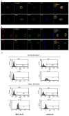
CXCR4 has the same cellular localization (cell surface and intracellular) in both cell lines. (A–B) Confocal micrographs of MOLT4 and Jurkat cell lines displaying CXCR7 (green) and CXCR4 (red) staining using 63× oil immersion objectives. Appropriated markers for membrane and cytoplasm were used to confirm the localization of these receptors: E-cadherin and Op18 present (yellow), respectively, in the membrane and in the cytoplasm. CXCR7 showed colocalization with these proteins in both cell lines; however CXCR7 was located mainly on the cell surface of MOLT4 cells; unlike, in Jurkat cells, where CXCR7 presented an intracellular and cell surface localization. CXCR4 had same cellular distribution (cell surface and intracellular) in both cell lines. (B) Flow Cytometry, a more quantitative method, confirmed the results observed in confocal microscopy because showed that less than 2% of MOLT4 cells versus 67% of Jurkat cells displayed intracellular CXCR7.
Figure 6
Figure 6. Lentivirus-mediated shRNA targeting CXCR7 effectively silenced CXCR7 in MOLT4 and Jurkat cells.
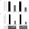
A) Quantitative expression of CXCR7 mRNA in cells relative to the shControl cells. mRNA expression levels of CXCR7 were normalized by HPRT and GAPDH endogenous control. Results were analyzed using 2−ΔΔCT. CXCR7 mRNA expression was reduced in MOLT4 cells (41%) and Jurkat cells (63%) when compared with shControl cells. (B) Western blotting analysis of shControl and shCXCR7 cell extracts. The membrane was blotted with antibodies against CXCR7 (42 kDa) or GAPDH (37 kDa), as a control for equal sample loading, and developed with the ECL Western Blot Analysis System. The bar graphs represent the band intensity of CXCR7 protein expression corrected for loading differences based on the corresponding GAPDH bands (UN-SCAN-IT software). Protein levels of CXCR7 were also reduced in MOLT4 cells (63%) and Jurkat cells (74%) when compared with shControl cells.
Figure 7
Figure 7. CXCR7 silencing decreases MOLT4 and Jurkat cell migration.
Cell migration toward either RPMI with 0.1% BSA and RPMI or 0.1% BSA containing CXCL12 (200 ng/mL) used as negative control and chemoattractant, respectively. After 4 h, the number of migrated cells was counted and was expressed as a percentage of the input, i.e., the number of cells applied directly to the lower compartment in parallel wells. The migration of cells was normalized to 100% +/− sd of triplicates. (A) The CXCR7 silencing resulted in significant changes in MOLT4 chemotactic response (P = 0.0159). The inhibition of CXCR4-dependent chemotaxis by its antagonist AMD3100 (1.25 µg/mL) promoted a similar effect (P = 0.0159). Moreover, the silencing of CXCR7 plus the treatment with AMD3100 exhibited a synergistic effect in cell chemotactic capacity (P = 0.0086). (B) The same effect was observed with Jurkat cells. The CXCR7 silencing (P = 0.0366) or the inhibition of CXCR4-dependent chemotaxis by its antagonist AMD3100 (P = 0.019) reduced Jurkat chemotactic response. The simultaneous silencing of CXCR7 and treatment with AMD3100 also exhibited a synergistic effect upon cell chemotactic capacity (P = 0.0191); Mann-Whitney test.
Figure 8
Figure 8. CXCR7 silencing did not modify apoptosis and proliferation of MOLT4 and Jurkat cells.
To evaluate whether CXCR7 is important in the process of cell death, control and inhibited CXCR7 cells were exposed to 10 J/m2 UV for different periods of time (0, 3, and 6 hours) and apoptosis was detected by flow cytometry using Annexin V/PI staining method. Cell proliferation was determined by MTT assay. Results are shown as mean ±SD of six replicates. No differences in apoptosis rate (A) or proliferation (B) were observed in MOLT4 and Jurkat cell lines.
Figure 9
Figure 9. High CXCR7 expression of peripheral blood and bone marrow lymphocytes.
(A) CXCR7 is expressed in peripheral blood leukocytes, however an increase in CXCR7 cell surface expression was observed in lymphocytes compared to monocytes and neutrophils. This difference was more apparent and significant when the cells were permeabilized (lymphocytes vs. monocytes, P = 0.0265 and lymphocytes vs. neutrophils, P = 0.0286) showing that the localization of this receptor is mainly intracellular in B-lymphocytes, CD4+ T-lymphocytes and CD8+ T-lymphocytes; Mann-Whitney test.

References

    1. Lodowski DT, Palczewski K (2009) Chemokine receptors and other G protein-coupled receptors. Curr Opin HIV AIDS 4: 88–95. - PMC - PubMed
    1. Mizoguchi T, Verkade H, Heath JK, Kuroiwa A, Kikuchi Y (2008) Sdf1/Cxcr4 signaling controls the dorsal migration of endodermal cells during zebrafish gastrulation. Development 135: 2521–2529. - PubMed
    1. Decaillot FM, Kazmi MA, Lin Y, Ray-Saha S, Sakmar TP, et al. (2011) CXCR7/CXCR4 heterodimer constitutively recruits beta-arrestin to enhance cell migration. J Biol Chem 286: 32188–32197. - PMC - PubMed
    1. Bachelerie F, Ben-Baruch A, Burkhardt AM, Combadiere C, Farber JM, et al. (2013) International Union of Pharmacology. LXXXIX. Update on the Extended Family of Chemokine Receptors and Introducing a New Nomenclature for Atypical Chemokine Receptors. Pharmacol Rev 66: 1–79. - PMC - PubMed
    1. Smart N, Riley PR (2008) The stem cell movement. Circ Res 102: 1155–1168. - PubMed

Publication types

MeSH terms